
Presentation
Heavy smoker.
Patient Data

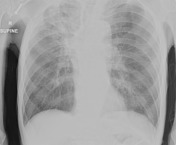
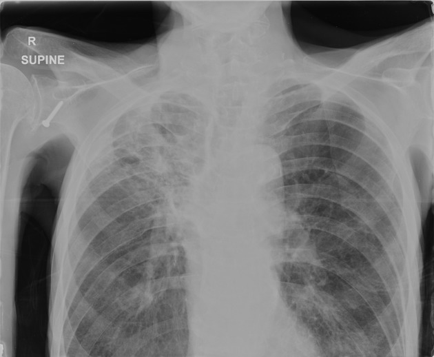
A spiculated opacity within the right upper lobe with surrounding extensive scarring and volume loss. Hyper-expansion of the lungs with distortion of the background architecture. Screw projects over the right glenoid.


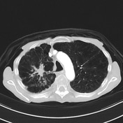
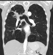
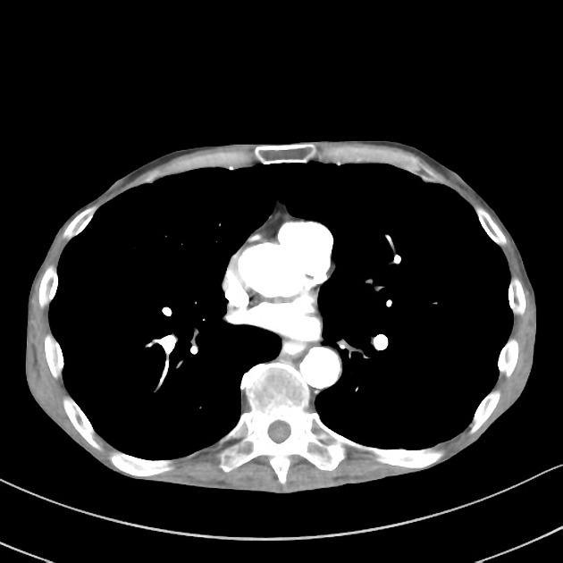
Right upper lobe spiculated mass resulting in narrowing of the right upper lobe bronchus. The spiculated mass. There is also a region of no mass-like consolidation in the apical segment distal to this which is likely post-obstructive infection/inflammation. There has been some improvement in left lower lobe inflammatory change. There are a few subcentimetre nodules persisting. Opacification of the proximal left lower lobe basal segmental bronchi in combination with contrast opacification of the oesophageal lumen, suggesting aspiration as the aetiology of these basal inflammatory changes. Scattered subcentimetre nodules in the right lung are stable in size. Background changes of severe emphysema are again noted. No pleural effusion. No thoracic lymphadenopathy.
Histology:
MICROSCOPIC DESCRIPTION: 1. The smears and cell block section contain scattered cohesive groups of malignant epithelial cells. The tumour cells have enlarged hyperchromatic nuclei, granular chromatin, inconspicuous nucleoli, nuclear moulding and scanty cytoplasm. Apoptotic debris, necrosis and some mitoses are present in the background. The tumour cells are synaptophysin, CD56 and TTF-1 positive. The features are those of small cell carcinoma.
DIAGNOSIS: 1. TBNA Right Hilar Mass: Small cell carcinoma.
Case Discussion
This case demonstrates a histologically proven lung small cell carcinoma in a background of lung emphysematous changes in a heavy smoker patient.


 Unable to process the form. Check for errors and try again.