Presentation
Elderly woman with a mass in the left lower abdomen with intense pain in this area for about three days but without nausea and vomiting. On physical examination, an ill-defined mass measuring about 4 cm is palpable, accompanied with high tenderness.
Patient Data
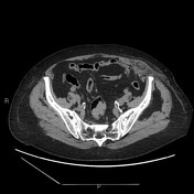

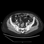

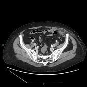


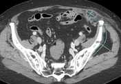
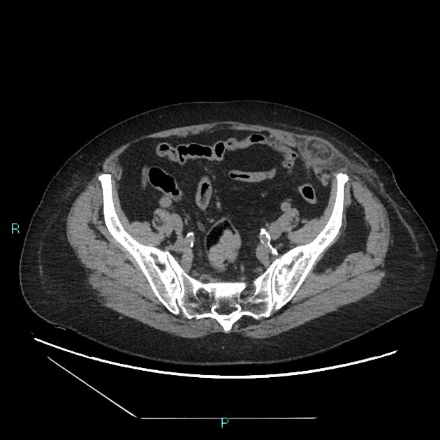
CT shows herniation of the omentum across the lateral border of the left rectus muscle in the lunate line with incarceration of preperitoneal fat that appears hyperdense consistent with ischemia. There is no peritoneal effusion or free air.
There is a 10 mm hepatic hemangioma at segment 5 and a tiny cyst at segment 8. It is also associated with uncomplicated diverticulosis of the descending colon and lipoma of the left gluteus medius muscle of about 10 cm.

Surgery report (Italian English translation)
Umbilical open laparoscopy and induction of pneumoperitoneum. Presence of left Spigelian hernia with part of the omentum incarcerated and necrotic, which is reduced and removed. Prosthesis repair.
Case Discussion
Spigelian hernia is the protrusion of properitoneal fat or peritoneal sac through a congenital or acquired defect in the Spigelian aponeurosis, in the aponeurotic layer between the rectus abdominis muscle medially and the lunate line laterally. Spigelian hernias usually contain only the small bowel or omentum.
Case courtesy of Dr.ssa Chiara Gennari
Radiographer: TSRM Fabio Imola


 Unable to process the form. Check for errors and try again.