
Presentation
Biopsy performed by renal team for investigation of declining renal function. The patient subsequently experienced severe left upper quadrant pain.
Patient Data
Age: 70 years
Gender: Male
From the case:
Splenic and pancreatic injury following renal biopsy


Download
Info
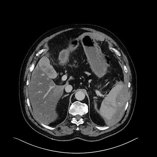
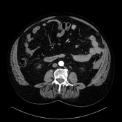
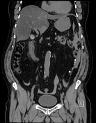
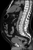
CT was performed 12 hours following the biopsy:
- There is a mixed-density crescentic collection indenting the lateral margin of the spleen, in keeping with a subcapsular hematoma.
- A hypodense tract can be seen extending through the spleen.
- On the arterial phase study, a tiny blush is evident within the posterior spleen, without expansion on the portal venous phase CT.
- Fluid is present within the anterior pararenal space and there is fatty stranding surrounding the pancreatic tail.
- Moderate volume high-density fluid (blood) within the pelvis and left paracolic gutter.
- Background hepatic steatosis. Small left pleural effusion.
The patient's serum lipase was elevated.
From the case:
Splenic and pancreatic injury following renal biopsy
Download
Info

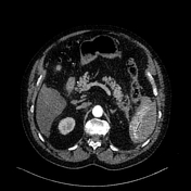
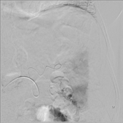
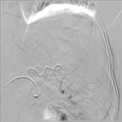
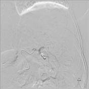
The patient's hemoglobin continued to reduce over the next 48hrs. He was referred to interventional radiology and a splenic artery angiogram was performed.
Three small pseudoaneurysms were identified. Microcoils were placed across a segmental splenic artery with good angiographic result.
Case Discussion
Renal hematoma is the most common complication following renal biopsy. Non-renal injuries can rarely occur.


 Unable to process the form. Check for errors and try again.