
Presentation
Patient referred from outside hospital with splenic lesion for evaluation.
Patient Data
Age: 30 years
Gender: Male
From the case:
Splenic hemangioma


Download
Info
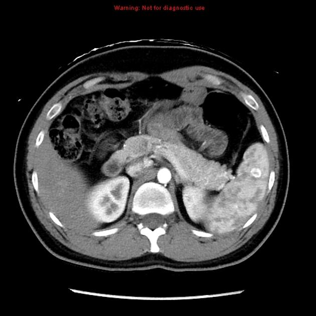
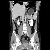
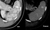
CT scan of the abdomen at the arterial phase reveals peripheral enhancement of the splenic lesion with subsequent fill-in in the delayed phases.
From the case:
Splenic hemangioma
Download
Info
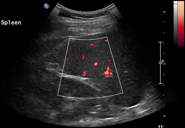
US abdomen performed and reveals a well defined hypoechoic lesion at the upper pole of the spleen without significant vascularity.
Case Discussion
The lesion is consistent with solitary splenic hemangioma.


 Unable to process the form. Check for errors and try again.