
Presentation
Severe left upper abdominal pain radiating across the abdomen and becoming less severe. Of course, the referral was to investigate epigastric/RUQ pain. She did not reveal any risk factors for arterial disease.
Patient Data

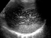
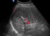
The spleen demonstrates a hypoechoic, hetrogeneous, wedge-shaped region characteristic of splenic infarction, extending across the entire width of the spleen. Doppler interrogation demonstrated absence of blood flow in the affected region.

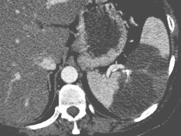
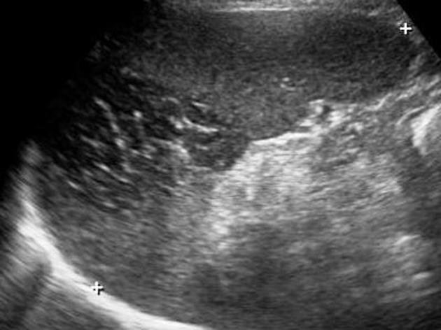
CT demonstrated a large region of altered attenuation within the spleen.
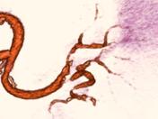
CT angiogram revealed multiple sites of stenosis and occlusion within the splenic artery branches.
- Note: This case has been tagged as "legacy" as it no longer meets image preparation and/or other case publication guidelines.
Case Discussion
The spleen demonstrated the classic ultrasound appearance of a splenic infarct: a hypoechoic, wedge-shaped defect. There was no evidence of other causes of the left-sided pain such as pancreatitis, inferior mesenteric artery stenosis or urinary tract pathology.
CT confirmed the infarction and CTA revealed significant disease within branches of the splenic artery.
The patient was referred to the local public hospital where she was counseled regarding total splenectomy. She declined this procedure. At a checkup 3 months after these examinations, she said the pain had disappeared.
CT images courtesy of Mr. David Sloane, radiographer, and Dr. Gary Geier, radiologist


 Unable to process the form. Check for errors and try again.