
Presentation
Right knee pain.
Patient Data
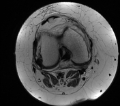

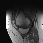
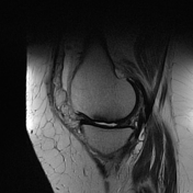
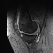

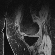


Subchondral T2 and PD hypointense crescent line is noted at medial femoral condyle with cortical surface irregularity and moderate surrounding marrow edema reflecting spontaneous osteonecrosis of the knee.
Complex meniscal tear is noted at the anterior horn lateral meniscus and also at the posterior horn medial meniscus. Grade I sprain of the medial collateral ligament.
Thinned out retropatellar and medial femorotibial cartilage with subchondral cysts reflecting osteoarthrosis. Mild joint effusion with synovitis. Mild subcutaneous edema around the knee. Semimembranosus bursitis is also noted.
Case Discussion
MRI features are suggestive of spontaneous osteonecrosis of the knee (SONK) involving the medial femoral condyle. It is usually a diagnosis in an older age group (>55 years) with a female predilection and usually unilateral, mostly affecting medial tibial plateau. This type of knee disease occurs with no history of trauma, however being considered as an insufficiency fracture on top of osteoporosis. It is often associated with a meniscal tear.
MRI suggestive features are:
- location: in the weight-bearing area, usually the medial femoral condyle
- subtle flattening / focal depressive deformity
- an irregular, discontinuous hypointense line in the subarticular marrow
- ill-defined bone marrow edema and lack of peripheral low signal intensity rim as seen in osteonecrosis and bone infarcts


 Unable to process the form. Check for errors and try again.