
Presentation
Acute difficulty breathing after a relatively short history of cough and temperature.
Patient Data
Age: 3 months
Gender: Male
From the case:
Spontaneous pneumothorax
Download
Info
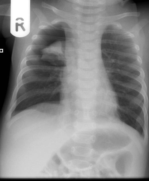
Focal opacification in the right upper zone with a region of hyperlucency peripherally. The peripheral zone does not have any bronchovascular markings. Features are of a pneumothorax complicating a chest infection (likely bronchiolitis).
Download
Info
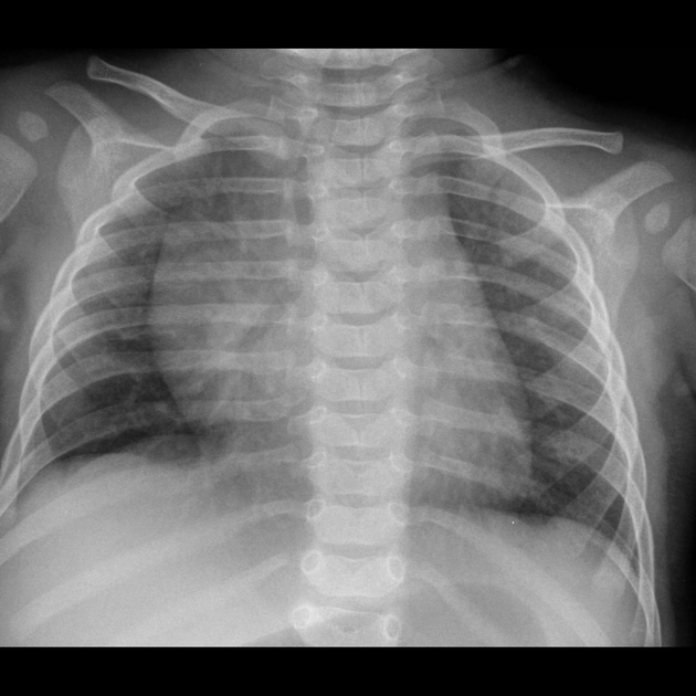
Following treatment with an intercostal drain and IV antibiotics, the chest x-ray is normal. No underlying cause.
Case Discussion
This 3 month old infant has a right-sided spontaneous pneumothorax. It was treated with intercostal drain insertion and after 3 months, there was complete resolution.
No features to suggest an underlying cause and no suggestion of NAI.


 Unable to process the form. Check for errors and try again.