
Presentation
Direct trauma to medial right clavicle
Patient Data

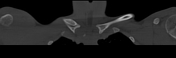
Superior dislocation of the right sternoclavicular joint is seen. In addition, there is a linear non- displaced fracture involving the medial clavicle.


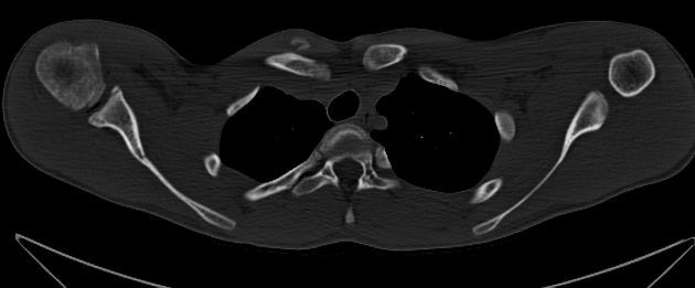
CT confirms a healing non displaced fracture involving the medial right clavicle and associated posterior dislocation of the ipsilateral sternoclavicular joint. The tip of the medial right clavicle lies between the braciocephalic trunk and superior vena cava. No hematoma, allowing for limitations of non contrast examination.
In addition, there is posterosuperior subluxation of left sternoclavicular joint. No fracture is identified on this side.
Case Discussion
The subsequent CT was performed 17 days after the clavicle radiograph, as the referring clinician suspected a left sided sternoclavicular dislocation on review of the patient. This explains the periosteal new bone formation over the medial clavicle, consistent with interval healing.


 Unable to process the form. Check for errors and try again.