
Presentation
Prostate cancer with PSA of 2492 ng/ml. Staging exam.
Patient Data


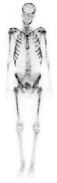
Single-phase whole-body bone scan in the anterior and posterior projections. Images were obtained approximately 3 hours after the IV administration of 25 mCi of Tc-99m MDP.
Images show abnormally intense radiotracer uptake throughout the axial and appendicular skeleton, consistent with widespread prostate metastases.
There is an abnormal absence of physiologic renal activity.
This combination is consistent with a superscan.
Case Discussion
A bone scan that demonstrates diffusely increased uptake of the radiopharmaceutical with absent or faint physiologic renal activity is known as a superscan.
The mechanism for a superscan is diffuse, increased osteoblastic activity, either due to widespread bony metastatic disease (most common) or a metabolic disease such as hyperparathyroidism (less common). Because of the markedly increased osseous uptake, the amount of unbound radiopharmaceutical decreases dramatically, and little is available for renal excretion; hence, the kidneys are either not visualized or only seen faintly.
The appearance of a metastatic superscan differs from that of a metabolic superscan. With metastatic disease (most commonly prostate or breast cancer), the uptake is heterogeneous or patchy and favors the axial skeleton. With metabolic bone disease, the increased uptake is often more homogeneous and may extend to the distal appendicular skeleton.


 Unable to process the form. Check for errors and try again.