
SVC thrombus, pulmonary embolism and myocardial infarction in LAD territory
Presentation
Follow up for known metastatic small bowel carcinoma.
Patient Data


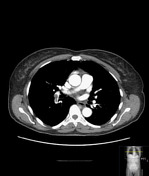
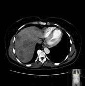
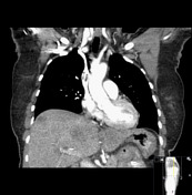
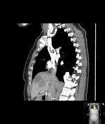
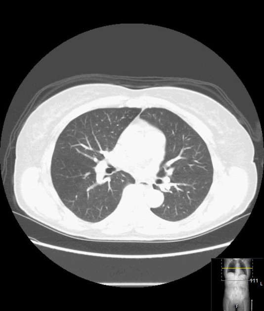
Central line in the SVC with adherent thrombus. Pulmonary embolism right interlobar artery. Decreased attenuation interventricular septum and left ventricular apex in the distribution of the left anterior descending artery. Multiple liver metastases, no intrathoracic metastases.
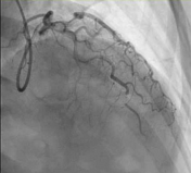

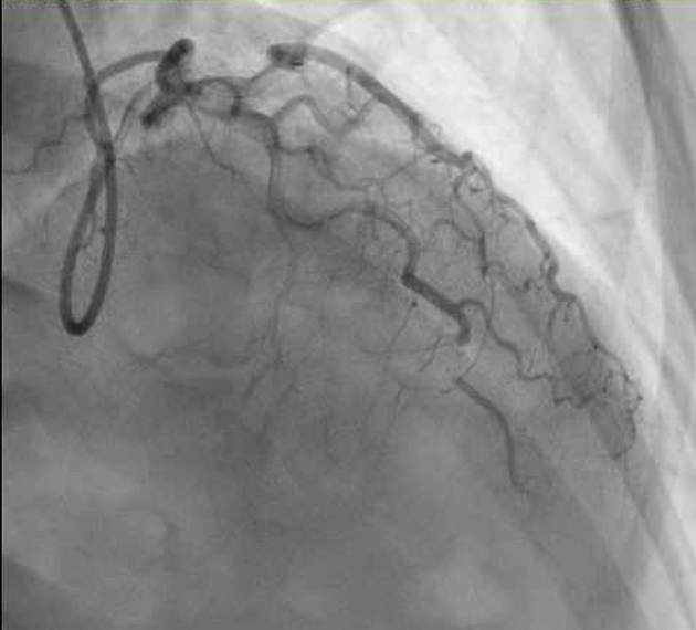
Occlusion distal LAD with distal reconstitution.
Case Discussion
This case illustrates the need to keep looking even if you find one, two or even three abnormalities. The metastases to the liver are easy to spot, but in addition, the patient had SVC thrombus, pulmonary embolism and findings of ischemia in the distribution of the LAD with distal LAD occlusion shown on subsequent coronary catheterization. Remarkably, the patient was asymptomatic.


 Unable to process the form. Check for errors and try again.