
Presentation
Minor head injury, with some minor chronic headaches.
Patient Data
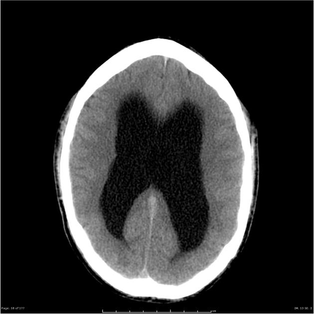
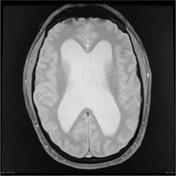
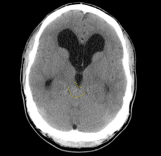
The lateral and third ventricles are markedly enlarged down to the level of the aqueduct. The tectum of the midbrain is expanded.

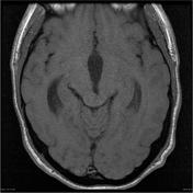
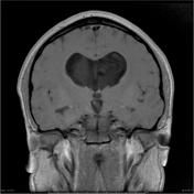


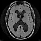
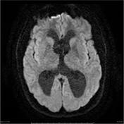

The right side of the tectal plate is expanded by a nodular mass which measures 15 x 14 x 11 millimeters in size. It is hyperintense on T2, isointense on T1, and demonstrates no contrast enhancement or restricted diffusion. The aqueduct is compressed with prominent obstructive hydrocephalus of the third and lateral ventricles. There is no transependymal edema, marked thinning of the corpus callosum, and fenestration of the septum pellucidum, which is cavum and demonstrates prominent flow artefact anteriorly.
The cerebellar tonsils are low lying, protruding 8 mm below foramen magnum in keeping with an acquired Chiari one malformation.
The remainder of the brain is unremarkable with no intra or extra-axial abnormality.

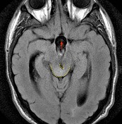
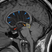
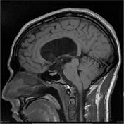
The tectum of the midbrain is expanded on the right by an ill defined mass (yellow dotted line) with resultant obstruction to the aqueduct (orange arrow).
There is hydrocephalus with marked upward deviation and thinning of the corpus callosum (blue arrows) and ballooning of the supraoptic recess fo the third ventricle (red arrow).
Case Discussion
Typical appearances of an indolent tectal plate glioma resulting in obstructive hydrocephalus. The patient went on to have a third ventriculostomy.


 Unable to process the form. Check for errors and try again.