
Presentation
Several episodes of seizures.
Patient Data
Age: 25 years
Gender: Female
From the case:
Temporal lobe cavernoma


Download
Info
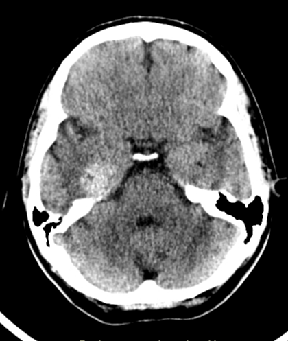
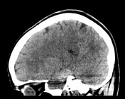
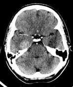
NECT of brain shows hyperdense ovoid lesion in right temporal lobe without perilesional edema. The lesion has no visible enhancement.
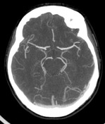
CTA demonstrates absence of brain AV malformations or any developmental venous anomaly (DVA).
From the case:
Temporal lobe cavernoma

Download
Info

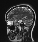
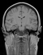
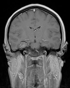
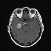
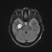
MRI demonstrates solitary bubbly-like lesion in right temporal lobe, with hypointense rim on T2, with minimal contrast enhancement (best seen on coronal T1). There are a prominent blooming on SWI which is compatible with hemosiderin. No perilesional edema.
Case Discussion
Classical CT and MRI picture of cavernoma of right temporal lobe.
Practical points: cavernoma looks like a hyperdense lesion without contrast enhancement on CT, has a prominent blooming on SWI/GRE T2*.


 Unable to process the form. Check for errors and try again.