
Presentation
Mass left hemiscrotum.
Patient Data
Age: 40 years
Gender: Male
From the case:
Testicular seminoma


Download
Info
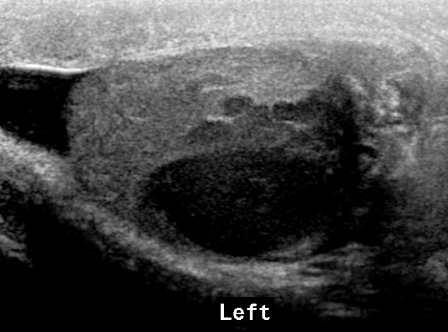
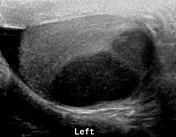
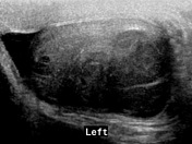
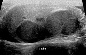
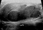
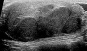
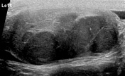
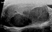
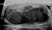
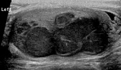
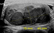
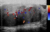
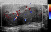
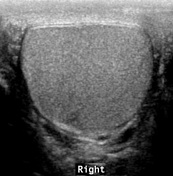
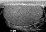
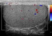
Multiple lobulated hypoechoic lesions, collectively measuring approximately 2 x 4 cm, are seen in the left testis. Few tiny calcifications are seen in these lesions. Color Doppler ultrasound examination shows increased vascularity in these lesions. Minimal left hydrocele. Right testis is normal.
Download
Info
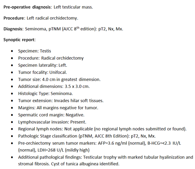
Histopathological analysis of the left orchiectomy specimen shows seminoma.
Case Discussion
Sonographic findings were suggestive of a multifocal malignant process affecting the left testis; however, the histopathology showed only unifocal disease.
The patient was referred to the medical oncologist for further evaluation/management.


 Unable to process the form. Check for errors and try again.