
Presentation
Chronic cough. Chest x-ray showed an anterior mediastinal mass.
Patient Data
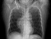


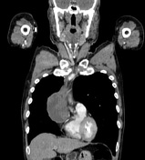

There is a smooth anterior mediastinal mass, with mixed internal density of containing both enhancing soft tissue and cystic areas. The outline of the mass is relatively well defined. No lymphadenopathy, pleural effusion or infiltration.
HISTOPATHOLOGY REPORT
MICROSCOPIC DESCRIPTION: Microscopy shows an encapsulated tumor composed of two alternating cytomorphological and histoarchitectural components. The first component contains neoplastic epithelium forming delicate loose network, and having large polygonal cells with large
nuclei, open chromating and prominent central nucleoli, equally admixed with non-neoplastic lymphocytes. The second component exhibits vaguely solid sheets of neoplastic epithelium composed of polygonal medium-sized cells, with smaller round nuclei and prominent nucleoli, with paucity of lymphocytes. Both components show abundance of Hassall's corpuscles. There are areas of extensive tumor necrosis, small cystic space formation and cholesterol clefts. There is no marked nuclear polymorphism or significant mitotic activity.
DIAGNOSIS: Corpuscular thymoma, WHO type B3.
Case Discussion
Thymomas are a common cause of anterior mediastinal masses.


 Unable to process the form. Check for errors and try again.