
Presentation
Rapidly growing neck mass
Patient Data
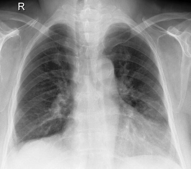
Deviation of the trachea to the right at the level of the thoracic inlet. Normal hila. Small right pleural effusion. Lungs clear.
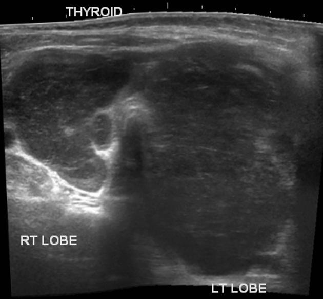
Enlarged thyroid gland is largely replaced by heterogeneous masses with marked hypoechogenicity. Neither significant vascularity nor calcification are seen. Multiple hypoechoic lymph nodes.
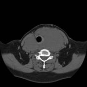


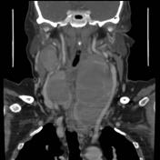
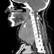

Precontrast images demonstrate loss of the inherit normal high density of the thyroid gland which is enlarged and replaced by hypodense masses. Post contrast study show enhancing septa within the non-enhancing enlarged thyroid. Multiple enlarged lymph nodes are seen as well.
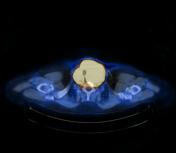
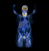
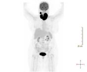
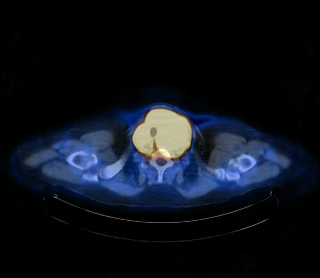
Marked activity within the enlarged thyroid gland. Small solitary active abdominal lymph node is also present of unclear significance.
Case Discussion
Pathologically-proven thyroid lymphoma.


 Unable to process the form. Check for errors and try again.