
Presentation
Stridor and associated neck mass.
Patient Data


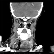
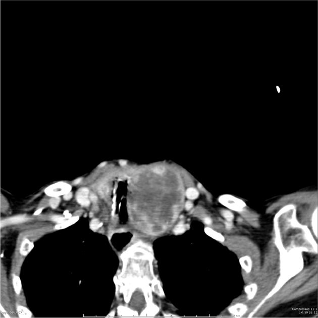
This is a limited study due to motion artefact. CT neck shows a large mass arising from left lobe of thyroid gland resulting in tracheal deviation and compression. The calcification of the trachea on the left is lost and there is extraluminal air consistent with direct invasion. There is also impression of further hypodense nodules within the right lobe of the thyroid gland.
Case Discussion
Patient underwent surgery to debulk thyroid gland and required a tracheostomy to maintain airway. Direct invasion was confirmed.
Histology
Sections through thyroid show thyroid which predominantly multinodular architecture with nodules separated by fibrous bands consisting of follicles of varying size. No intranuclear halos, grooves or psammoma bodies are seen. However on the posterior surface of the thyroid and infiltrating into the thyroid there are focal areas of a diffuse population of large atypical haematolymphoid cells which have large nuclei and a moderate amount of eosinophilic cytoplasm. There is a focal area which shows these large atypical cells showing angiocentric invasion. These atypical cells only form a small proportion of the thyroid. Immunohistochemistry show these cells stain strongly positive with Cd20. Negative with Cd5, Cd10, Cyclin D1.
Conclusion:
Multinodular adenomatous goiter with focal area of diffuse large B cell lymphoma, focally within thyroid and extending beyond thyroid.


 Unable to process the form. Check for errors and try again.