
Presentation
Heavy smoker complaining of an ulcerated lesion on his tongue.
Patient Data
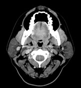

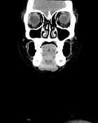
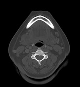

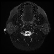
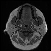
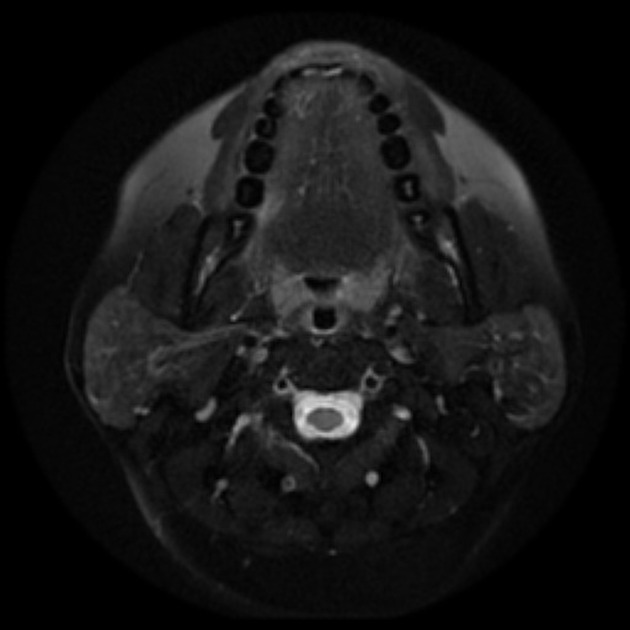
There is an enhancing plaque like lesion at the right lateral posterior margin of the oral tongue. This measures 7 x 14.6 x 12.6 mm (TR/AP/CC).
There is no definite involvement of the extrinsic muscles of the tongue although the tumor is close to the superior margin of the right hyoglossus muscle. There are number of cervical lymph nodes but none of which is suspicious by size or imaging criteria.
No destructive osseous lesion.
Conclusion: Right lateral tongue SCC (suggested radiological staging T1 N0).
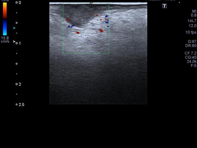
At the site of the right posterolateral tongue, there is a focal, well-defined hypoechoic lesion which has a maximum depth perpendicular to the tongue lateral surface 6 mm. It measures 12 mm in diameter at the surface of the tongue.


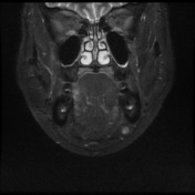

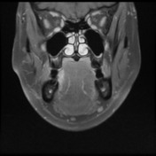
Focal lesion is seen in the right lateral tongue, in the posterior aspect of the oral tongue. Maximum depth is 6.3 millimeters. The length of the lesion is 10 mm. No clear extrinsic muscle involvement.
Mildly prominent bilateral submandibular and cervical chain lymph nodes, greater on the left.
Conclusion: MR findings suspicious for a right lateral oral tongue carcinoma, maximum depth 6.3 millimeters.
Case Discussion
The case was confirmed as a squamous cell carcinoma of the tongue. This tumor has tobacco smoking and alcohol ingestion as major risk factors. These tumors usually arise from the ventrolateral aspect of the mid and posterior tongue.
Whether CT or MRI is used these features should be assessed:
- size of tumor and tumor thickness
- extension across the midline
- extension beyond the intrinsic muscles of the tongue
- involvement of adjacent structures
- neurovascular bundle and submandibular duct in the floor of mouth
- mandible


 Unable to process the form. Check for errors and try again.