
Presentation
Wrist trauma
Patient Data
Age: 20 years
Gender: Male
From the case:
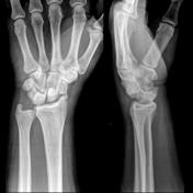
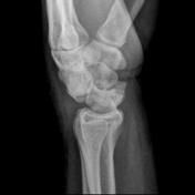
Trans-scaphoid perilunate dislocation

Download
Info
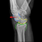
Fracture of the scaphoid with dorsal dislocation of the carpus relative to the lunate.
From the case:
Trans-scaphoid perilunate dislocation
Download
Info
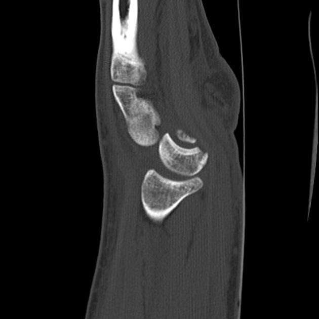
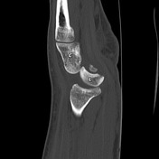
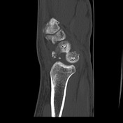
CT confirms the findings seen on the plain films, and better delineates the multiple small fragments at the scaphoid fracture. Note how the radiolunate joint is a little widened but remains aligned.
From the case:
Trans-scaphoid perilunate dislocation
Download
Info
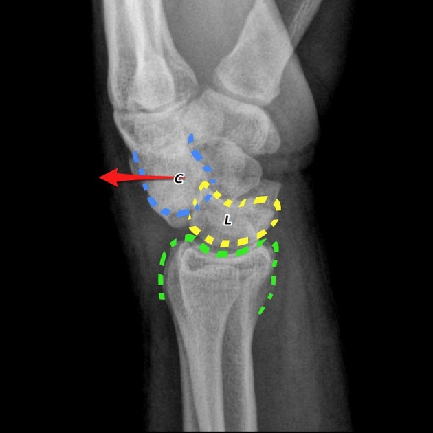
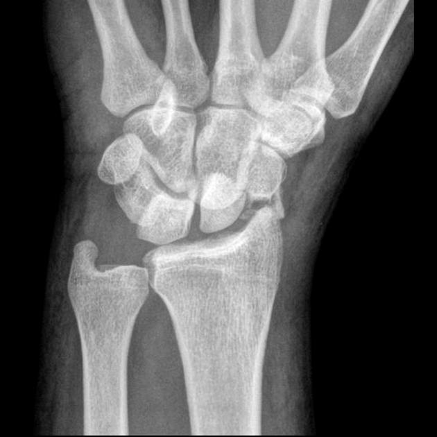
Capitate (C, blue) is dislocated dorsal to the lunate (L, yellow). The proximal pole of the scaphoid (PP) has remained aligned with the lunate, whereas the distal pole (DP) has followed the capitate dorsally. The lunate and the distal radius (green) remain normally aligned.
Case Discussion
Typical appearances of a trans-scaphoid perilunate dislocation.


 Unable to process the form. Check for errors and try again.