
Presentation
Head trauma in a road traffic collision.
Patient Data
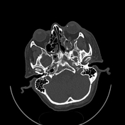

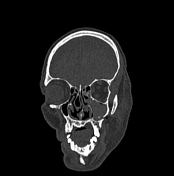
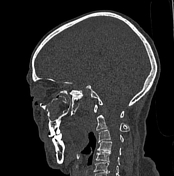

Multiple fractures in face and cranial bones:
left zygomatic arch, with intact temporozygomatic suture
posterior wall of maxillary sinus, lateral wall of ethmoid sinus, and left lateral pterygoid plate; associated with zygomaticomaxillary suture diastasis
fracture of inferior orbital rim (blowout fracture) and medial aspect of orbit (lamina papyracea fracture); associated with diastasis of frontozygomatic suture
patent optic nerve canals, however, a tiny hyperdense focus is noted at left globe sclera (?bone fragment)
right-sided concha bullosa (incidental finding anatomical variant)
Case Discussion
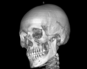
The features of multiple traumatic left sided facial fractures.
A zygomaticomaxillary complex fracture, also known as a tripod fracture, is a complex injury involving multiple bones. This type of fracture extends to multiple nearby facial cavities, including the orbit, the paranasal sinuses, and the mandibular ramus.
It requires sophisticated internal fixation by maxillofacial surgery, as the cheekbone can alter the shape of the face.
Careful assessment of the optic nerve canals and pathways is essential to evaluate vision preservation.


 Unable to process the form. Check for errors and try again.