
Presentation
Growth delay.
Patient Data


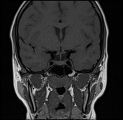
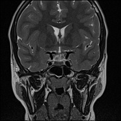
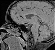
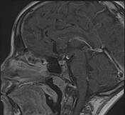
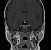
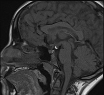
A well-defined, non-enhancing fat-containing lesion is observed in the tuber cinereum region, located between the mammillary bodies and the pituitary stalk.
The lesion appears hyperintense on T1 and T2 weighted images and suppresses on fat-saturation sequences, consistent with a lipoma.
There is no abnormality of the pituitary gland and its stalk.
The posterior pituitary is in its normal position, with a normal high signal on T1.
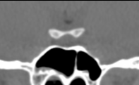
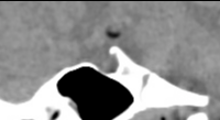
Complementary CT confirms the fat density of the lesion with no evidence of calcification.
Case Discussion
A tuber cinereum lipoma is a rare, benign congenital lesion composed of mature fat, often discovered incidentally on brain imaging like in our patient. Differentiating it from other hypothalamic lesions is crucial. Hypothalamic hamartomas are iso- to hypointense on T1 and do not suppress on fat-saturation sequences. Dermoid cysts, though also fat-containing, may have heterogeneous signals due to sebaceous material and may contain calcifications. Recognizing these imaging features helps avoid unnecessary investigations or interventions.


 Unable to process the form. Check for errors and try again.