Presentation
The patient presented to the emergency department with symptoms of headache, nausea, and vomiting, followed by the onset of disorientation, neck stiffness, fever, and a decline in clinical condition, requiring orotracheal intubation and intensive care therapy. Furthermore, there is a reported history of previous diagnoses and irregular treatment for pulmonary tuberculosis.
Patient Data
![](https://prod-images-static.radiopaedia.org/images/63071083/003a04aee25755d0028c262b5b49b2c0b274c1e69f592db4e740cf5592f5f20e_thumb.jpeg)
![This study is a stack](/packs/stack-YQKLCKBI.gif)
![](https://prod-images-static.radiopaedia.org/images/63071160/9acef6c397ac13413541cdd042da97507a691ac322ddbbaa754e26d13de2542a_thumb.jpeg)
![This study is a stack](/packs/stack-YQKLCKBI.gif)
![](https://prod-images-static.radiopaedia.org/images/63071083/003a04aee25755d0028c262b5b49b2c0b274c1e69f592db4e740cf5592f5f20e_big_gallery.jpeg)
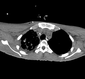
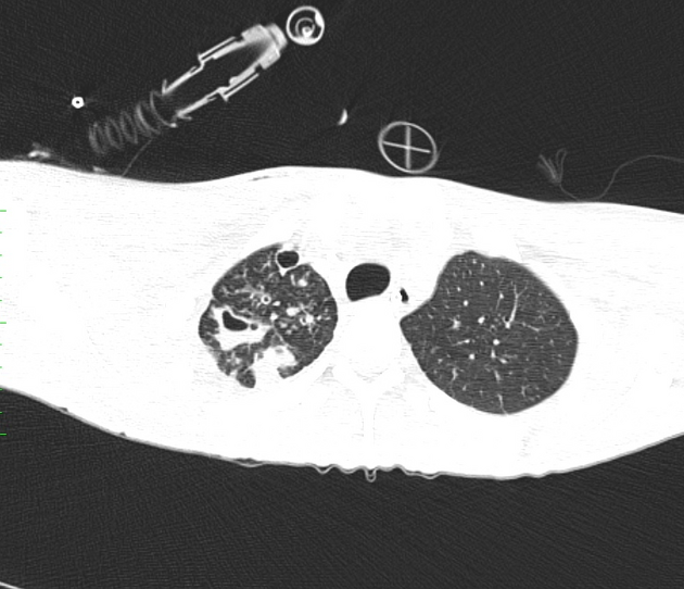
Non-enhanced chest computed tomography (CT) shows thick-walled cavities, bronchiectasis, and some patchy areas of consolidation in the right lung, with an endobronchial spread characterized by centrilobular nodules and tree-in-bud pattern. Right pleural effusion.
Impression: The findings are in keeping with pulmonary tuberculosis.
![](https://prod-images-static.radiopaedia.org/images/63071647/7b2e2e9a65f18291c85cc33ca80fbfeffca652c12b0ee26e572a43d127eabee2_thumb.jpeg)
![This study is a stack](/packs/stack-YQKLCKBI.gif)
![](https://prod-images-static.radiopaedia.org/images/63071748/b67552fa034569bcabe9a8e283919bbe5c7fba5d1a46c699021b5d1bac17ae54_thumb.jpeg)
![This study is a stack](/packs/stack-YQKLCKBI.gif)
![](https://prod-images-static.radiopaedia.org/images/63071940/457aa4146c489645249c30ed4e8ba88cbe37140c6059dcd7503684d844e18f33_thumb.jpeg)
![This study is a stack](/packs/stack-YQKLCKBI.gif)
![](https://prod-images-static.radiopaedia.org/images/63072001/f86827712c9fbf976f21659692e46293714940b27e53e7267f7b22f6fe329882_thumb.jpeg)
![This study is a stack](/packs/stack-YQKLCKBI.gif)
![](https://prod-images-static.radiopaedia.org/images/63072056/eadc6352f771b58626382ec0a946bd0d1ea0771dba6c9088bac66243f78014a7_thumb.jpeg)
![This study is a stack](/packs/stack-YQKLCKBI.gif)
![](https://prod-images-static.radiopaedia.org/images/63072083/6c7e3b06c31516bdd2a2b3469a3bd2136633c73d506b02392865279ed416ffc7_thumb.jpeg)
![This study is a stack](/packs/stack-YQKLCKBI.gif)
![](https://prod-images-static.radiopaedia.org/images/63072133/1862449c9e7646d19a0fc9ecd252e1deedbcd6f62ce7d49b93ff802225b9e202_thumb.jpeg)
![This study is a stack](/packs/stack-YQKLCKBI.gif)
![](https://prod-images-static.radiopaedia.org/images/63072162/496a7ba5bbe2bade5f1660f7de68db2a4ff805dba210d51240fb67aeeb824fc2_thumb.jpeg)
![This study is a stack](/packs/stack-YQKLCKBI.gif)
![](https://prod-images-static.radiopaedia.org/images/63072193/4ee4b15c4cd53626873b0358a756de17cf4f11241c3c5a07095feb6c4de406d3_thumb.jpeg)
![This study is a stack](/packs/stack-YQKLCKBI.gif)
![](https://prod-images-static.radiopaedia.org/images/63072223/947bcf12aaaf9ef74d2bafff01ed7d54fb77110a87dbaf6d0b0d8e8a17fa65ec_thumb.jpeg)
![This study is a stack](/packs/stack-YQKLCKBI.gif)
![](https://prod-images-static.radiopaedia.org/images/63071647/7b2e2e9a65f18291c85cc33ca80fbfeffca652c12b0ee26e572a43d127eabee2_big_gallery.jpeg)
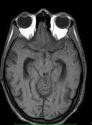
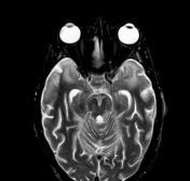
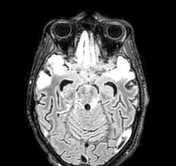
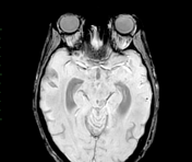
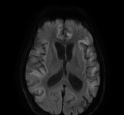
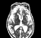
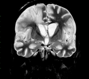
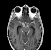
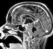
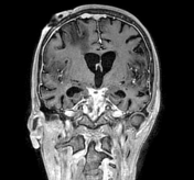
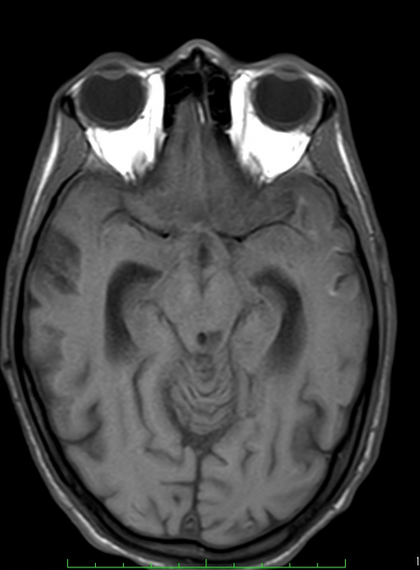
Magnetic resonance imaging (MRI) reveals altered signal areas in the bilateral perisylvian cortical-subcortical region, with subtle restricted diffusion on DWI. There are enhancing exudates at the basal cisterns and bilateral thickened leptomeningeal enhancement, most pronounced at the basal cisterns, and some degree of involvement of the meninges in the Sylvian fissures and within the sulci over the cerebral convexities. There is a left thalamic well-defined chronic infarction T1 hypointense and T2 hyperintense lesion without diffusion restriction and no enhancement. A right-sided ventricular shunt catheter was in situ because a previous CT revealed hydrocephalus.
Impression: Features are consistent with tuberculous meningitis.
Case Discussion
Pulmonary tuberculosis, a contagious mycobacterial infection, can disseminate, leading to tuberculous meningitis, a severe life-threatening disease affecting the meninges 1-4. Early diagnosis, treatment, and diligent infection control measures are crucial in managing both conditions effectively 1-4. This case aims alert radiologists about the relationship between pulmonary tuberculosis and tuberculous meningitis.
Case courtesy
Murilo Pessoa, MD - PGY-2, radiology resident, Department of Radiology
Fabiano Ferreira, MD - PGY-2, neurology resident, Department of Neurology
Antonio Rodrigues de Aguiar Neto, MD - radiologist, Department of Radiology
Hospital da Restauração – Recife, PE – Brazil