Presentation
Two months history of Headaches and low-grade fever with recent episodes of seizures and altered mental status.
Patient Data
![](https://prod-images-static.radiopaedia.org/images/51866278/440d3fe8263138f3fe19f08402e396_thumb.jpeg)
![This study is a stack](/packs/stack-YQKLCKBI.gif)
![](https://prod-images-static.radiopaedia.org/images/51866307/e6598f4d00af52cf364ff47d44e16a_thumb.jpeg)
![This study is a stack](/packs/stack-YQKLCKBI.gif)
![](https://prod-images-static.radiopaedia.org/images/51866330/92def104b201a4781782009c098527_thumb.jpeg)
![This study is a stack](/packs/stack-YQKLCKBI.gif)
![](https://prod-images-static.radiopaedia.org/images/51866383/34b9ddbb24b70a63176c6d64252ee4_thumb.jpeg)
![This study is a stack](/packs/stack-YQKLCKBI.gif)
![](https://prod-images-static.radiopaedia.org/images/51866414/8fea70a2272418c13c02502b3e976c_thumb.jpeg)
![This study is a stack](/packs/stack-YQKLCKBI.gif)
![](https://prod-images-static.radiopaedia.org/images/51866434/610215990f33c517ab155cb19c4571_thumb.jpeg)
![This study is a stack](/packs/stack-YQKLCKBI.gif)
![](https://prod-images-static.radiopaedia.org/images/51866446/29107b70c39aebb25f03a7cdf77a2f_thumb.jpeg)
![This study is a stack](/packs/stack-YQKLCKBI.gif)
![](https://prod-images-static.radiopaedia.org/images/51866457/b27d71a6efb51878a9807de35612fa_thumb.jpeg)
![This study is a stack](/packs/stack-YQKLCKBI.gif)
![](https://prod-images-static.radiopaedia.org/images/51866278/440d3fe8263138f3fe19f08402e396_big_gallery.jpeg)
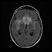
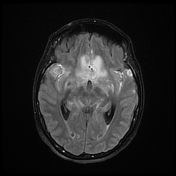
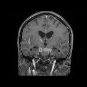
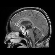
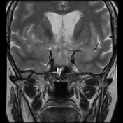
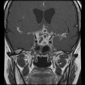
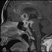
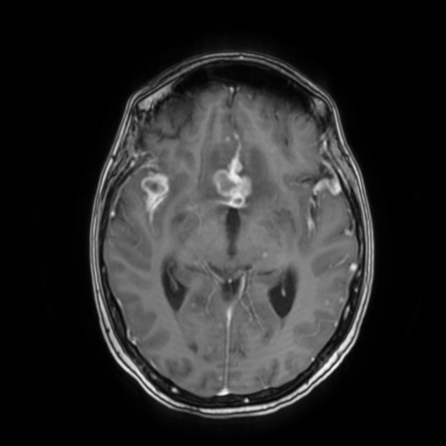
There is an extensive thickening of the basal subarachnoid cisterns with an intense enhancement, extending along the middle cerebral arteries to the Sylvian fissures, sellar and suprasellar regions as well as the cavernous sinuses with numerous conglomerate enhancing exudates and ring lesions (largest at the suprasellar region and right Sylvian fissure). Dilated 3rd, and lateral ventricles with mild interstitial periventricular edema, indicating obstructive hydrocephalus.
Extensive nodular and ring-like enhancement along the surface of the cerebellar and cerebral hemispheres as well as the bulb and brainstem.
Mild dilatation of the 3rd and lateral ventricles suggesting associated obstructive hydrocephalus.
![](https://prod-images-static.radiopaedia.org/images/51866623/7119746ca7ad8069d010dbdd616bb9_thumb.jpeg)
![This study is a stack](/packs/stack-YQKLCKBI.gif)
![](https://prod-images-static.radiopaedia.org/images/51866642/28fd3b48032c142dbd0267e97c7be1_thumb.jpeg)
![This study is a stack](/packs/stack-YQKLCKBI.gif)
![](https://prod-images-static.radiopaedia.org/images/51866525/d264fe8954c26b428cb060afdcc0ea_thumb.jpeg)
![This study is a stack](/packs/stack-YQKLCKBI.gif)
![](https://prod-images-static.radiopaedia.org/images/51866623/7119746ca7ad8069d010dbdd616bb9_big_gallery.jpeg)
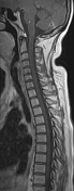
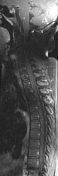
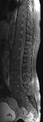
Extensive linear and nodular pial thickening with enhancement along the dorsal, and ventral aspect of the entire spinal cord up to the cauda equina.
Case Discussion
The MRI findings are suggestive of tuberculous meningitis with cerebral tuberculomas and spinal subarachnoid involvement.
The CSF analysis with culture was positive (tuberculous bacilli)