
Presentation
Pulsatile tinnnitis. Retrotympanic reddish mass.
Patient Data
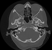


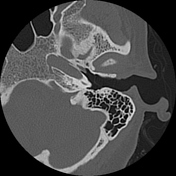
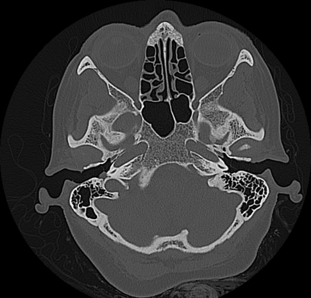
A retrotympanic small soft tissue density lesion is noted at the right middle ear cavity, lateral to the promontry, oval window and facial recess. Intact jugular plate and floor of tympanic cavity.

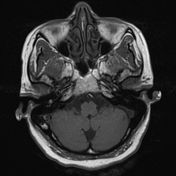
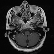
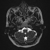
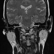
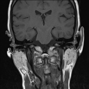
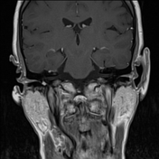
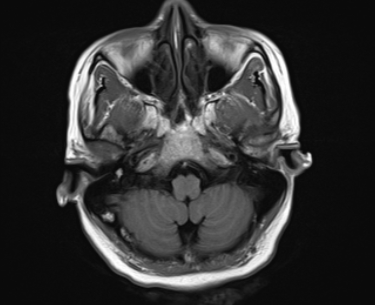
Right middle ear cavity shows a 6 mm lobular opacity is noted at the region of promontory, round window and facial recess, showing diffuse post-gadolinium enhancement. Intact jugular plate and floor of tympanic cavity.
Case Discussion
Right tympanic paraganglioma (Glasscock/Jackson - type 1) 1. Tympanic paragangliomas are the most common tumor of the middle ear cavity. They arise from the Jacobson nerve at the cochlear promontory. It appears as a soft tissue mass lateral to the cochlear promontory with intact jugular bulb. Contrast-enhanced MRI shows avid enhancement.
Case courtesy Prof. Dr Mohamed Eid, Professor of Radiodiagnosis, Alexandria University, Egypt.


 Unable to process the form. Check for errors and try again.