
Presentation
Chronic diarrhea.
Patient Data
Age: 25 years
Gender: Female
From the case:
Ulcerative colitis


Download
Info

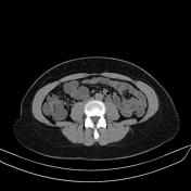
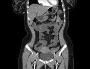
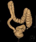
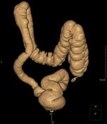
CT enterography study revealed circumferential mural thickening and mucosal enhancement of the rectum, sigmoid and descending colon with dilated perisigmoid vasculature and hyperattenuation of the adjacent fat.
Normal CT enterography study of the small bowel loops.
Two tiny (segment VIII & IV) hepatic cysts.
Right renal cortical cyst showing fine enhancing internal septae (Bosniak II)
Small uterine fibroid.
From the case:
Ulcerative colitis

Download
Info
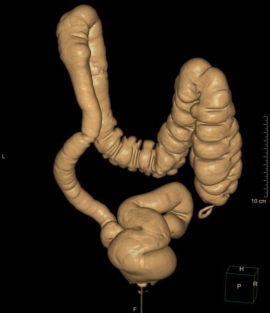
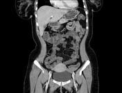
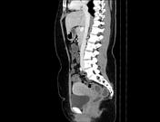
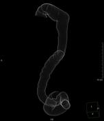
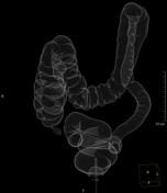
CT colonoscopy study revealed loss of haustral pattern in the rectum, sigmoid and descending colon giving the "lead pipe sign".
Case Discussion
This case shows the appearance of ulcerative colitis at both CT enterography and CT colonography studies.
Diagnosis was confirmed by endoscopy.


 Unable to process the form. Check for errors and try again.