
Presentation
Deteriorating renal function. Cause unknown.
Patient Data
Note: This case has been tagged as "legacy" as it no longer meets image preparation and/or other case publication guidelines.
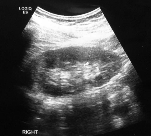
Diagnostic ultrasound image prior to biopsy
Normal BPD and cortical thickness.
The width of the cortex is measured over the lower pole prior to biopsy, to gauge positioning of the needle tip, allowing for the 2cm throw of the biopsy needle.
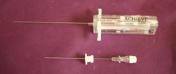


Co-axial needle for when more than one core is taken. 18G cores are typically taken.
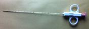
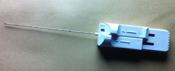
Other biopsy devices shown.

Dynamic image as biopsy taken.
The core biopsy aims to taken the renal cortex, without significant medullary fat, and certainly avoiding the pelviocalyceal system.
Case Discussion
Ultrasound-guided renal biopsy may be performed for:
non-focal biopsy of the native kidney to investigate the cause of renal impairment
focal biopsy for an unclassified lesion on diagnostic imaging
non-focal biopsy of a transplanted kidney to investigate the cause of renal impairment


 Unable to process the form. Check for errors and try again.