
Presentation
Progressive abdominal distension and palpable mass in right upper quadrant for about 6 months, associated with considerable weight loss. Prior fine-needle biopsy concluded extensive necrosis of infectious etiology.
Patient Data
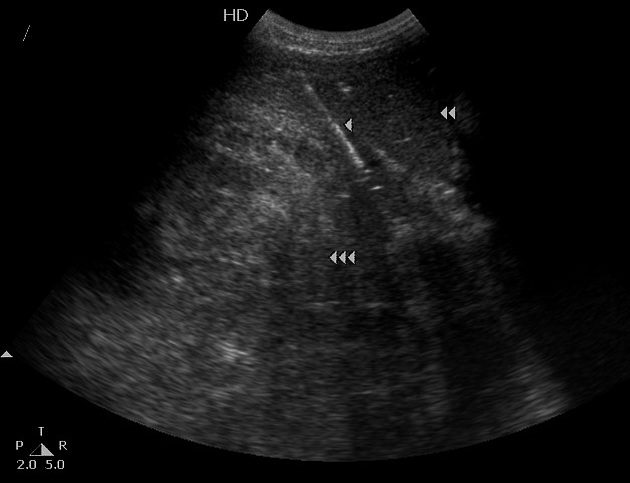
Abdominal ultrasound-guided biopsy shows an extensive, ill-defined, heterogeneous mass (triple arrowhead) located in the right liver lobe, with an apparently normal left liver lobe (double arrowhead). The biopsy needle is shown by the single arrowhead.


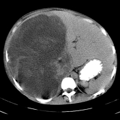
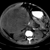
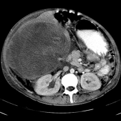
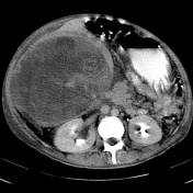
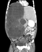
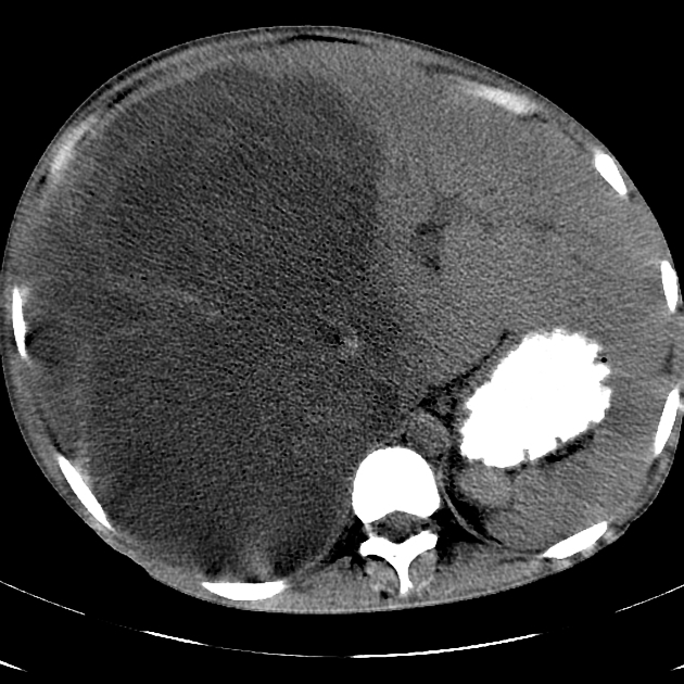
The abdominal CT study shows an extensive hypodense mass centered in the right liver, with an estimated volume over 7,000 cc. The mass contains thick septa and solid nodular elements, and results in right hydronephrosis due to ureteral compression. The C+ delayed phase shows slight enhancement of the septa and nodular elements. Retroperitoneal and mesenteric lymph nodes were detected. No evidence of portal venous thrombosis was seen.
Case Discussion
This patient was initially evaluated for a possible infectious disease, due to the initial biopsy findings. However, no proof of an infective agent was found after multiple laboratory tests. Alpha-fetoprotein (AFP) levels were in normal range.
A repeat US-guided biopsy revealed anaplastic fusiform cells with hyperchromatic multiple nuclei and a myxoid matrix associated with extensive necrosis, highly consistent with embryonal sarcoma of the liver.
This pathology is considered by some authors as the malignant counterpart of hepatic mesenchymal hamartoma.


 Unable to process the form. Check for errors and try again.