
Presentation
Two week history of fevers and right upper quadrant pain.
Patient Data


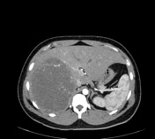
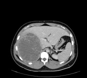
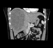
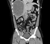
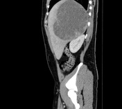
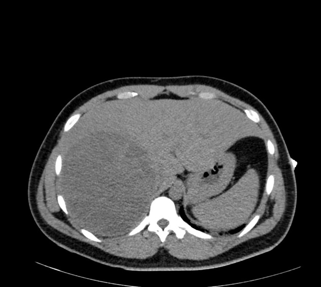
Large low density liver mass with enhancing septations.
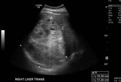
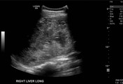
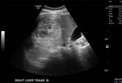

A large heterogeneous lesion is noted in the right lobe of the liver. This is hyperechoic relative to the background liver, was thought to perhaps contain cystic spaces.
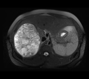
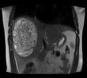

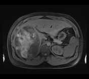


A large hepatic lesion is again noted, with heterogeneous high T2 signal intensity relative to the background liver. The lesion contains multiple low T2 signal bands, likely fibrous septae.
Heterogeneous high T1 internal signal is present in the lesion pre-contrast, suggestive of hemorrhage. Minor peripheral gadolinium contrast-enhancement is present.
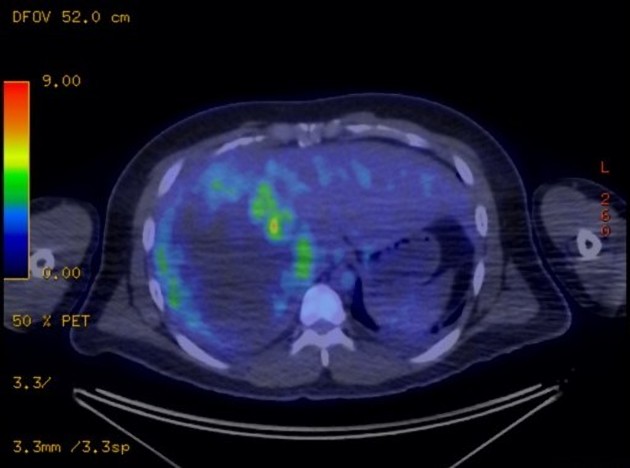
There is abnormal FDG avidity around the periphery of the large low density lesion in the right lobe of liver. Based solely on FDG PET, a distinction between a large liver abscess and a predominantly necrotic primary or secondary liver tumor cannot be made.
Histology Report
Embryonal sarcoma, with evidence of lymphovascular invasion.
Case Discussion
The patient proceeded to a partial hepatectomy, partial adrenalectomy and resection of the diaphragm. A biopsy was not performed.
Hepatic embryonal sarcoma is a rare tumor in the adult population.


 Unable to process the form. Check for errors and try again.