
Presentation
Diffuse abdominal pain.
Patient Data


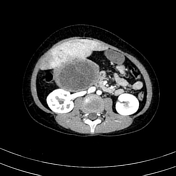
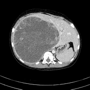
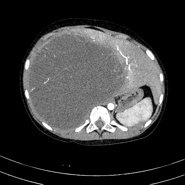
Large mass (152 x 153 x 185 mm) located in the right lobe of the liver, with solid and non-enhancing large fluid attenuation components.
The lesion is hypervascular and heterogeneous enhancement. Pseudocapsule and internal multiple septa show progress enhancement in portal venous and delayed contrast phase. The septa are more dominant around the periphery of the mass.
Significant mass effect to the right kidney and pancreatic head. No IVC or portal vein thrombus.
The left liver is unremarkable.
Case Discussion
Righ lobe liver mass with typical CT appearances of undifferentiated embryonal sarcoma:
- pediatric patient under 15 years
- location: right lobe of the liver
- large mass
- significant necrotic component
- multiple septa showing delay phase enhancement
The patient underwent a biopsy. Histology revealed undifferentiated embryonal sarcoma.
Undifferentiated embryonal sarcomas of the liver are rare, aggressive liver tumors encountered in the pediatric population. The prognosis has historically been poor.


 Unable to process the form. Check for errors and try again.