
Presentation
Intermittent abdominal pain for past few weeks, increasing abdominal girth.
Patient Data
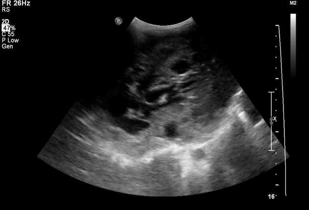
Hepatic mass measuring 10 cm in diameter involving most of the liver, with locules of fluid, some of which contain turbid fluid.
The gallbladder cannot be demonstrated. The bile ducts are not dilated.
Vascular interrogation cannot be completed due to (understandable) lack of cooperation.
Small amount of pelvic fluid with mobile sediment, most probably exudate.
Normal appearing spleen, kidneys, and urinary bladder.
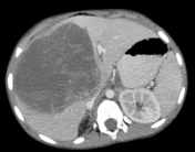

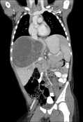
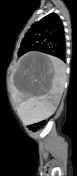

CT chest-abdomen-pelvis done on same day as the US:
Huge hypodense, heterogeneous mass in the center of the liver measuring 11 x 12.5 x 12 cm, containing locules of fluid-fluid levels and blood vessels. No calcification. The mass medially displaces all components of the hepatic hilum and compresses the right portal branch. Middle and right hepatic veins involved, lateral tributary of right hepatic vein normal. Rest of liver shows heterogeneous texture secondary to vascular compromise. The IVC is compressed at the caudate lobe level, without visible luminal invasion. Soft tissue fullness at root of mesentery around mesenteric vessels, as well as low attenuation retroperitoneal lymph nodes arounf the aorta and the IVC. Periportal edema.
Non-dilated bile ducts, gallbladder normal.
Minute pleural effusion, right greater than left.
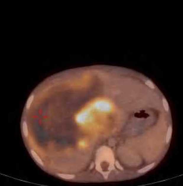
PET-CT done 1 week later:
Enlarged liver with huge well-defined hypodense, heterogeneous hepatic mass measuring 12.8x13x14 cm, occupying most of the right lobe. Heterogeneous, predominantly peripheral metabolic uptake, and in the solid-appearing areas in the medial part of the mass. The mass has enlarged cf. the CT study - intralesional bleeding?
No additional pathologic hepatic findings or abnormal uptake elsewhere.
Bilateral pleural effusion with no pathologic uptake, has enlarged cf. the CT study. Passive RLL atelectasis.
Patent portal vein.
Hypodense periportal changes denoting edema.
Free intra-abdominal fluid around the liver, in the right paracolic gutter, and small amount in the pelvis - new finding.
Diffuse bone marrow uptake, secondary to anemia.
Case Discussion
Brought by his parents due to a new abdominal distension, having complained of intermittent abdominal pain that would wake him from sleep. Systemic fever up to 38.5°C. 2-Kg weight loss during the past 2 weeks.
At the ER, he looked pale. The liver was palpated 4 cm below the costal arch. Right upper quadrant tenderness elicited. A single soft axillary node was palpated on the right.
Lab exams remarkable for elevated hepatocellular enzymes and normocytic hypochromic anemia.
Ultrasound demonstrated a huge hepatic mass, as did the CT and PET-CT scans. The mass was 30% larger on the PET-CT study, attributed to intralesional hemorrhage. This was verified on a repeat US (not shown).
Alpha fetoprotein 2.2 ng/mL (normal), CMV IgG positive.
Histopathology report of a biopsy obtained from the mass:
Tiny fragments from a liver mass, compatible with a malignant mesenchymal tumor.
A port was inserted and the patient was started on chemotherapy.
Histopathology report from the right lobectomy specimen:
Small foci represent less than 2% of the mass volume of residual undifferentiated sarcoma composed of large pleomorphic stromal cells, present at the edge of the tumor, focally infiltrate the sinusoids and stain very weakly and scattered cells positive for CD31 and desmin. KER MNF116, CD34, SMA, myogenin, and ERG negative in the pleomorphic cells.
The mass shows extensive treatment effect, with hyalinosis, abundant siderophages, necrosis, and residual bile ducts, thick walled vessels, and groups of hepatocytes mixed with pleomorphic cells.
India-inked surgical margin free of tumor in the sections examined. India-inked margins of the diaphragm shows fragment of muscle focally necrotic and tissue with marked treated artifacts. It is not possible to define residual tumor in these areas. Liver parenchyma preserved. Gallbladder unremarkable.
In light of the finding of pleomorphic cells and in light of the immunohistochemical assay results, the remnants are compatible with undifferentiated embryonal sarcoma.


 Unable to process the form. Check for errors and try again.