
Presentation
Disseminated cocci with CNS involvement complicated by hydrocephalus.
Patient Data


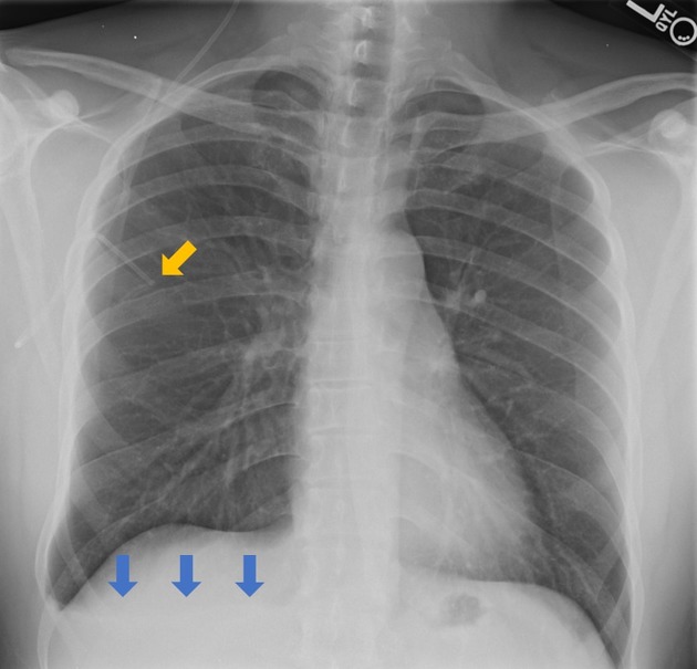
A cerebral shunt is seen tunneling within subcutaneous soft tissue and terminating in the right upper chest (orange arrow). Fluid layering is present in the right lung base consistent with a pleural effusion (blue arrows).
Case Discussion
Cerebral shunts are used to relieve intracranial pressure from hydrocephalus. Most shunts terminate in the peritoneum, however, other alternative destinations include the pleura, right atrium, or gallbladder. Causes of ventriculoperitoneal shunt placement requiring conversion include intraabdominal infection, peritonitis, pseudocyst formation, or obstruction. Complications of ventriculopleural shunts include pleural effusion and pneumothorax.
This case was submitted with supervision and input from:
Soni C. Chawla, M.D.
Associate Professor
Department of Radiological Sciences
David Geffen School of Medicine at UCLA
Olive View - UCLA Medical Center


 Unable to process the form. Check for errors and try again.