
Presentation
Left-sided renal colic.
Patient Data
Age: 40 years
Gender: Male
From the case:
Vesico-ureteric junction stone with resultant hydronephrosis
Download
Info
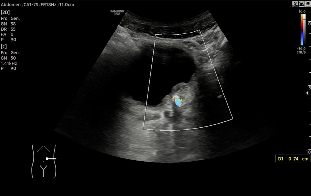
- Moderate left renal hydronephosis.
- Dilated (6-7 mm) left ureter which can be followed to the level of the iliac arteries.
- Impacted 7-8 mm stone in the left VUJ demonstrating twinkling.
Case Discussion
In patients with acute renal colic the region of the VUJ must be scrutinised for reflective stones. While this stone is fairly large and thus easily appreciated on B-mode imaging, for small stones the use of color Doppler and search for twinkling can enhance sensitivity. Waiting for color jets to appear in the bladder can also be used as an indirect way of confirming obstruction but in some patients no jets will appear even during prolonged observation regardless of the presence/absence of ureteric obstruction.


 Unable to process the form. Check for errors and try again.