
Presentation
Neurological abnormalities with vomiting. There was a query of septo-optic dysplasia on CT brain imaging.
Patient Data
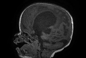


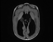
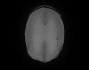

Agyria with smooth cortex and subcortical nodularity in keeping with cobblestone lissencephaly.
Severe hydrocephalus with absent septum pellucidum.
Dorsal kink at the mesencephalic-pontine junction resulting in a Z-configuration of the brainstem.
Hypoplastic vermis, and multiple bilateral cerebellar parenchymal cysts.
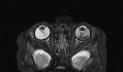
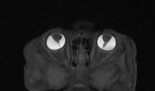
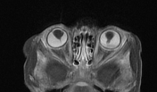
Bilateral persistent hyperplastic primary vitreous as evidenced by retrolental soft tissue with a stalk giving a martini glass-shape.
There is tractional retinal detachment on the left with layering debris in the vitreous.
There is thinning of the optic chiasm and bilateral optic nerves.
Case Discussion
Unfortunately, in this case, neither a muscle biopsy nor genetic tests were available. The presumptive diagnosis in this instance is based on the clinical and typical radiological characteristics of Walker-Warburg syndrome which include hydrocephalus, diffuse cobblestone lissencephaly, dorsal kink at the mesencephalic-pontine junction, cerebellar innumerable small cysts and ocular anomalies in the form of bilateral PHPV. A differential diagnosis of retinal detachment with hemorrhage is entertained since the retrolental stalk of soft tissue did not enhance, despite the imaging appearance resembling persistent hyperplastic primary vitreous.
Walker-Warburg syndrome (WWS) is a rare and severe genetic disorder characterized by a combination of brain, eye, and muscle abnormalities. It is the most severe form of congenital muscular dystrophy and is typically associated with a poor prognosis, often leading to death within the first few years of life.
Case co-contributors:
Dr Gary Peiser, Department of Radiology, Nelson Mandela Children Hospital, South Africa
Dr Nonceba Koranteng, Department of Radiology, Nelson Mandela Children Hospital, South Africa
Dr Gopolang Mndebele, Department of Radiology, Nelson Mandela Children Hospital, South Africa


 Unable to process the form. Check for errors and try again.