Presentation
Acute frank hematuria in the setting of longstanding mild abdominal pain
Patient Data

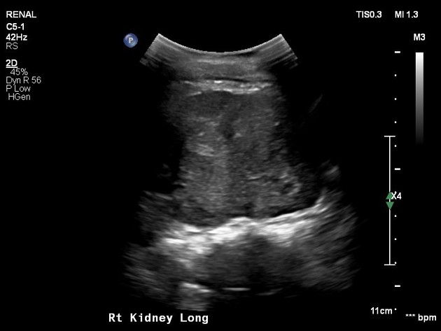
Solid heterogeneous mass occupying the mid to lower pole of the right kidney. It appears lobulated with the mid pole component measuring about 54x48x45mm and the lower pole exophytic component measuring at 50x49x41mm. The lesion is heterogeneous with significant internal vascularity. In the visualized segment, the renal vein appears patent but IVC could not be well evaluated. The upper pole collecting system is not grossly dilated.


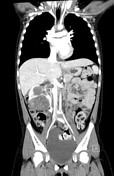
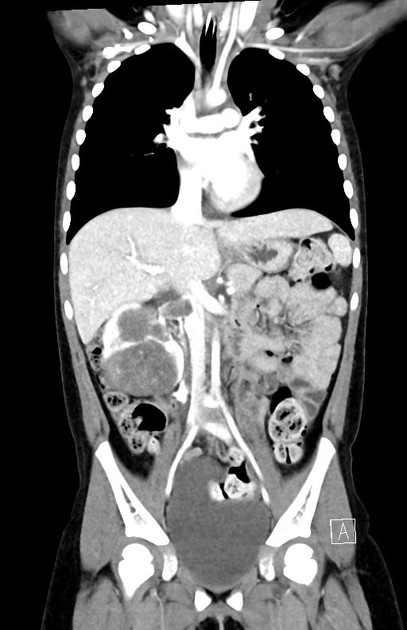
Large right renal tumor (6cm AP x 5.3cm TR x 6.8cm CC), with tumor thrombus extension into the right renal vein and IVC at the level of renal vein insertion.
Case Discussion
Given the patient’s age, the top differential diagnosis is Wilms tumor.
An ultrasound-guided biopsy performed show a classical triphasic Wilms tumor, with stromal, blastemal and epithelial components all present. No anaplasia is seen in this biopsy.


 Unable to process the form. Check for errors and try again.