
Presentation
Right flank pain with a palpable mass.
Patient Data
Age: 5 years
Gender: Female
From the case:
Wilms tumor


Download
Info

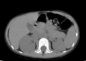
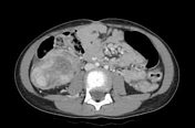
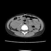
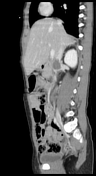
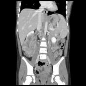
Heterogeneously enhancing exophytic soft tissue mass of the right kidney centered on the lower pole, invading the renal sinus with capsular effraction and infiltration of the perirenal fat with thickening of the anterior renal fascia. Thrombosis of the right renal vein and the adjacent segment of the IVC.
The tumor appears in contact with the hepatic flexure and ascending colon with effacement of the fatty interface. Minimal effusion is noted in the right paracolic gutter.
No evidence of nodal or distant abdominal metastatic disease.
Case Discussion
CT features of a large soft tissue mass of the right kidney invading the renal vein and IVC most consistent with Wilms tumor.


 Unable to process the form. Check for errors and try again.