
Radiation-induced MRI signal changes in bone marrow are the earliest detectable changes in bone. Their severity correlates with increasing radiation dose.
Pathology
1st week: decreased marrow cellularity with edema and hemorrhage
2nd week: increased marrow cellularity due to influx from non-irradiated areas
1-3 months: marrow sinusoids and cellularity is decreased
after 6 months: regeneration of hematopoietic tissue and sinusoids
Radiographic features
MRI
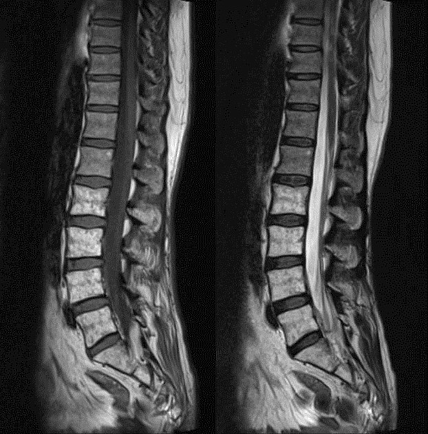
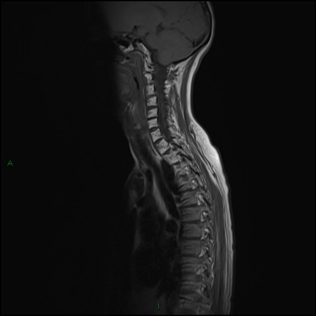
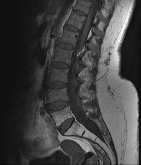
Little changes in signal intensity during the first 3 weeks of radiation therapy on spin echo sequences. STIR shows increased signal intensity during this period.
After 3 weeks heterogenous mottled appearance of marrow is seen.
During 3-6 weeks marrow cellularity is replaced by central fat predominantly surrounding the basivertebral veins.


 Unable to process the form. Check for errors and try again.