
Aggressive angiomyxomas are rare tumors that arise in the pelvis and typically cross the levator ani muscles. Despite its name, it is essentially a benign tumor and the term "aggressive" is due to a predilection for local recurrence. Only rarely does it metastasize.
On this page:
Epidemiology
It is seen predominantly in women (>90%) in the second through fourth decades of life 3.
Pathology
Aggressive angiomyxoma is a mesenchymal tumor that arises from connective tissue.
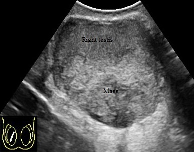
Deep angiomyxoma is seen in the vulva, vagina, perineum, inguinal area and pelvic soft tissue. It can be seen in men's shaft of the penis. The tumor appears as a slow growing mass and has a rubbery or gelatinous consistency.
Microscopically: spindle or stellate cells with bland nuclei and small inconspicuous nucleoli are embedded in a loose myxoid stroma with delicate collagen fibers. The mitotic count is very low and nuclear atypia is absent. There are various caliber dilated blood vessels accompanied by red blood cell extravasation in the stroma.
Deep angiomyxoma is particularly hypocellular and lacks necrosis.
IHC markers: Positive for desmin , SMA (smooth muscle actin) , MSA (muscle specific actin) , HMGA2 , CD34, ER, PR. Negative for S100, caldesmon and MDM2.
Molecular: Rearrangement of HMGA2 in one–third of aggressive angiomyxomas.
Local recurrence in approximately 30% of patients is reported but metastasis is uncommon. 5
Radiographic features
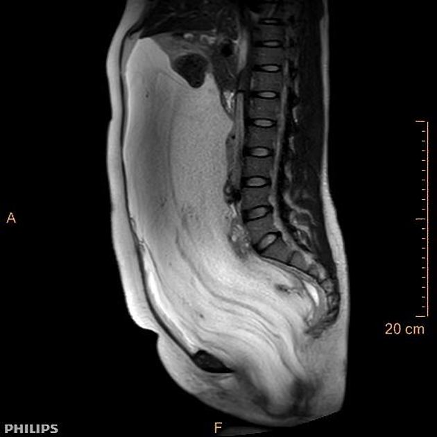
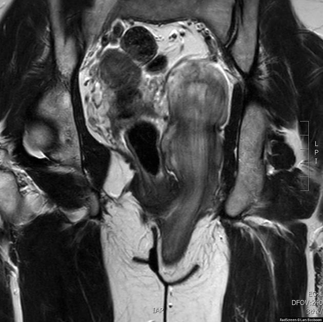
Classically shows involvement of both pelvic and perineal compartments. In males, it may involve the inguinoscrotal region 4.
CT
On CT, it usually presents as a well-defined mass with attenuation less than or equal to that of adjacent skeletal muscle. It often has swirled enhancing tissue internally 3.
MRI
Reported signal characteristics of the lesion include:
T1: tends to be iso to low signal
T2: predominantly high signal and typically has a swirled appearance
Due to the presence of collagen fibrils in the myxoid tissue, a laminated pattern may be seen on T2W images and post contrast T1 images, with alternating hyper- and hypointense linear areas 4. Rarely, cystic degeneration and intratumoral vessels may be seen 4.
Treatment and prognosis
Surgical resection with long term follow up is the treatment of choice. The "aggressive" tumor is infiltrative, so chances of local recurrence are high 4.
In ER- and PR-positive cases, hormonal therapy is also found to be helpful 4.


 Unable to process the form. Check for errors and try again.