
The blood-brain barrier (BBB) forms a physical resistance to the passage of lipophobic substances from cerebral capillaries into the brain and is a key reason why there is no CSF enhancement following intravenous contrast media on CT and MRI.
On this page:
Images:
Gross anatomy
The blood-brain barrier is formed by a combination of endothelial cells, pericytes, and astroglial and perivascular macrophages along the cerebral capillary walls.
In general, capillary walls in the human body can consist of three different types:
-
continuous: present in areas which have a blood-brain barrier
continuous interendothelial tight junctions
no pinocytosis
no fenestrations
fenestrated: present in areas which lack the blood-brain barrier
sinusoidal: not found in the brain
In the brain, the majority of capillary walls are of the continuous type, with tight junctions and a continuous basement membrane
Areas which contain fenestrated capillaries, and thus lack the blood-brain barrier, are:
Generally, lipophilic solutes can cross the blood-brain barrier, including:
Hydrophilic solutes, in general, are unable to cross it, e.g. water soluble CT/MRI contrast media.
Related pathology
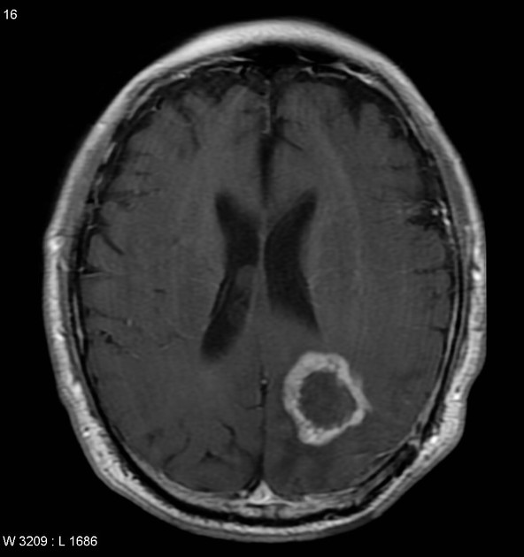
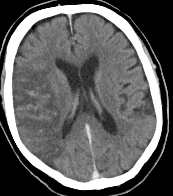
There are a multitude of conditions associated with disruption of the blood-brain barrier:
hypoxia, ischemia and infarction
tumors
inflammatory conditions, e.g. meningitis
trauma
intracranial irradiation
progressive multifocal leukoencephalopathy: the JC virus can cross the blood-brain barrier


 Unable to process the form. Check for errors and try again.