Gaillard F, Campos A, Sharma R, et al. Cerebral microhemorrhage. Reference article, Radiopaedia.org (Accessed on 13 Mar 2025) https://doi.org/10.53347/rID-4560
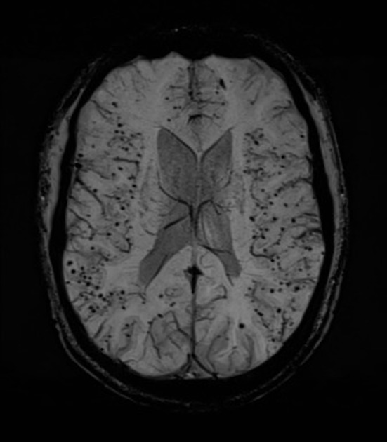
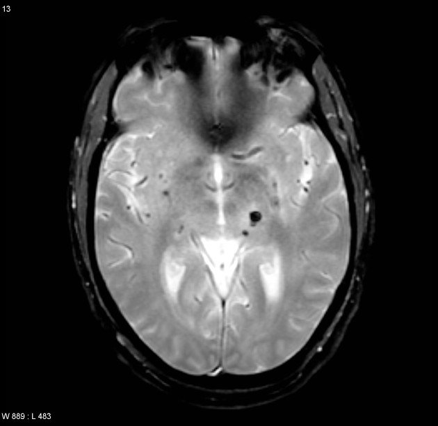
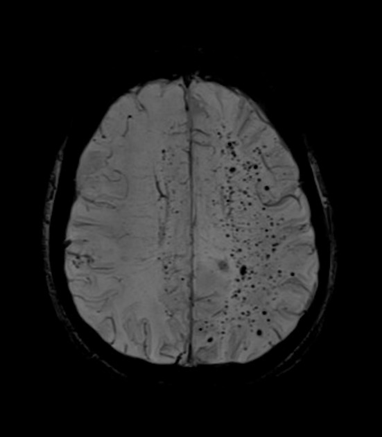
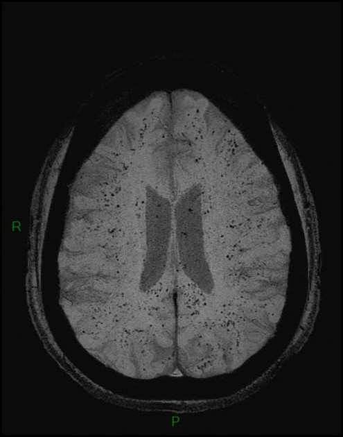
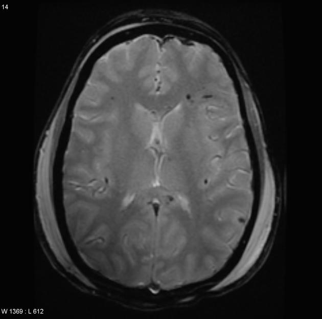
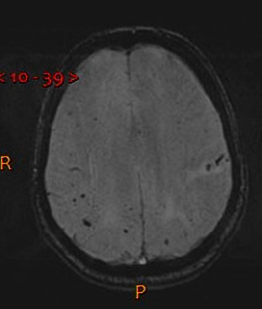
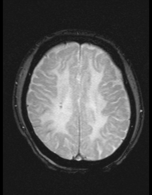
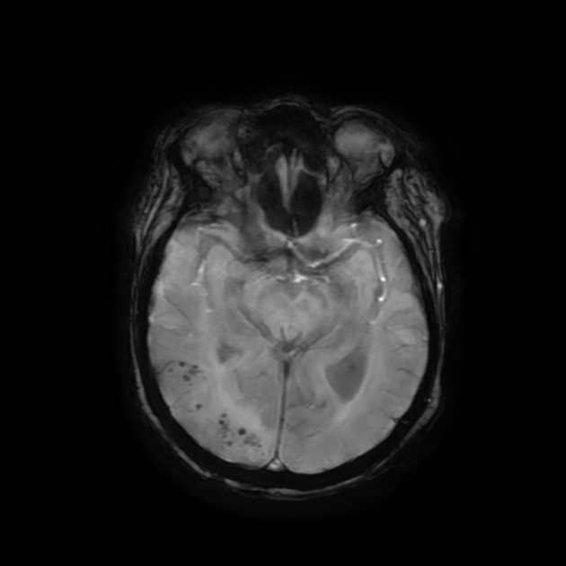
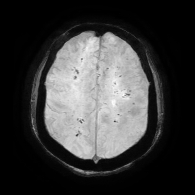
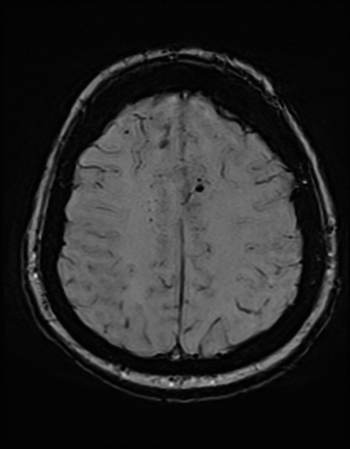
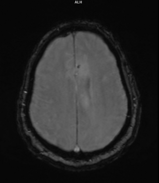
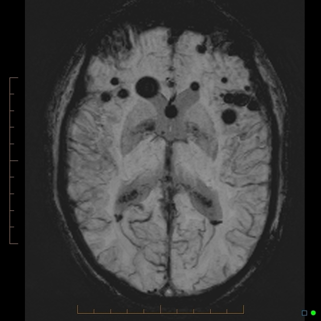
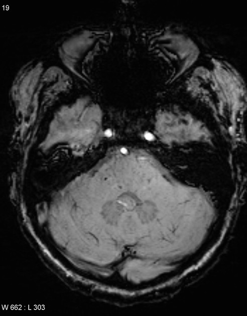
Cerebral microhemorrhages, or cerebral microbleeds,are small focal intracerebral hemorrhages, often only visible on susceptibility-sensitive MRI sequences.
They appear as conspicuous 2-10 mm punctate regions of signal drop out with blooming artifact24. This blooming grossly overestimates the size of the lesions, thus they are usually inapparent on other MRI sequences and CT 24.
4. Zaitsu Y, Terae S, Kudo K et-al. Susceptibility-weighted imaging of cerebral fat embolism. J Comput Assist Tomogr. 2010;34 (1): 107-12. doi:10.1097/RCT.0b013e3181a962c1 - Pubmed citation
5. Lanfranconi S, Markus HS. COL4A1 Mutations as a Monogenic Cause of Cerebral Small Vessel Disease. Stroke. 41 (8): e513. doi:10.1161/STROKEAHA.110.581918 - Pubmed
6. Jeon SB, Kang DW. Neurological picture. Cerebral air emboli on T2-weighted gradient-echo magnetic resonance imaging. Journal of neurology, neurosurgery, and psychiatry. 78 (8): 871. doi:10.1136/jnnp.2006.102954 - Pubmed
7. Liebeskind DS, Sanossian N, Sapo ML, Saver JL. Cerebral microbleeds after use of extracorporeal membrane oxygenation in children. Journal of neuroimaging : official journal of the American Society of Neuroimaging. 23 (1): 75-8. doi:10.1111/j.1552-6569.2012.00723.x - Pubmed
8. Sharma R, Dearaugo S, Infeld B, O'Sullivan R, Gerraty RP. Cerebral amyloid angiopathy: Review of clinico-radiological features and mimics. (2018) Journal of medical imaging and radiation oncology. doi:10.1111/1754-9485.12726 - Pubmed
10. Radmanesh A, Derman A, Lui Y et al. COVID-19-Associated Diffuse Leukoencephalopathy and Microhemorrhages. Radiology. 2020;297(1):E223-7. doi:10.1148/radiol.2020202040 - Pubmed
11. Igarashi S, Ando T, Takahashi T et al. Development of Cerebral Microbleeds in Patients with Cerebral Hyperperfusion Following Carotid Endarterectomy and Its Relation to Postoperative Cognitive Decline. J Neurosurg. 2021;135(4):1122-8. doi:10.3171/2020.7.jns202353 - Pubmed
12. Bathla G, Watal P, Gupta S, Nagpal P, Mohan S, Moritani T. Cerebrovascular Manifestations of Neurosarcoidosis: An Underrecognized Aspect of the Imaging Spectrum. AJNR Am J Neuroradiol. 2018;39(7):1194-200. doi:10.3174/ajnr.A5492 - Pubmed
13. Kammeyer R & Schreiner T. Cortical Vein Thrombosis, Tortuous Venous Vasculature, and Microhemorrhages in Neurosarcoidosis. JAMA Neurol. 2021;78(4):491. doi:10.1001/jamaneurol.2020.5440
14. Giyab O, Balogh B, Bogner P, Gergely O, Tóth A. Microbleeds Show a Characteristic Distribution in Cerebral Fat Embolism. Insights Imaging. 2021;12(1):42. doi:10.1186/s13244-021-00988-6 - Pubmed
15. Patel N, Banahan C, Janus J et al. Perioperative Cerebral Microbleeds After Adult Cardiac Surgery. Stroke. 2019;50(2):336-43. doi:10.1161/strokeaha.118.023355 - Pubmed
16. Sperling R, Jack C, Black S et al. Amyloid-Related Imaging Abnormalities in Amyloid-Modifying Therapeutic Trials: Recommendations from the Alzheimer's Association Research Roundtable Workgroup. Alzheimers Dement. 2011;7(4):367-85. doi:10.1016/j.jalz.2011.05.2351 - Pubmed
17. Richie M, Guterman E, Shah M, Cha S. Susceptibility-Weighted Imaging of Intravascular Lymphoma of the Central Nervous System. JAMA Neurol. 2022;79(1):86-7. doi:10.1001/jamaneurol.2021.4391 - Pubmed
18. Llufriu S, Cervera A, Capurro S, Chamorro A. Neurological Picture. Familial Sneddon's Syndrome with Microbleeds in MRI. J Neurol Neurosurg Psychiatry. 2008;79(8):962. doi:10.1136/jnnp.2007.131912 - Pubmed
19. Yao M, Zhao J, Jiang N, Li L, Ni J. Superficial Siderosis and Microbleed Restricted in Cortex Might Be Correlated to Atrophy and Cognitive Decline in Sneddon's Syndrome. Front Neurol. 2020;11:547600. doi:10.3389/fneur.2020.01035
20. Wen L, Yuan J, Li S et al. Case Report: Diffuse Cerebral Microbleeds in Cerebral Autosomal Recessive Arteriopathy With Subcortical Infarcts and Leukoencephalopathy. Front Neurol. 2022;13:818332. doi:10.3389/fneur.2022.818332 - Pubmed
21. Nozaki H, Sekine Y, Fukutake T et al. Characteristic Features and Progression of Abnormalities on MRI for CARASIL. Neurology. 2015;85(5):459-63. doi:10.1212/WNL.0000000000001803 - Pubmed
22. Mori N, Miki Y, Kikuta K et al. Microbleeds in Moyamoya Disease: Susceptibility-Weighted Imaging Versus T2*-Weighted Imaging at 3 Tesla. Invest Radiol. 2008;43(8):574-9. doi:10.1097/RLI.0b013e31817fb432 - Pubmed
23. Khan N, Saherwala A, Chen M et al. Prevalence of and Risk Factors for Cerebral Microbleeds in Moyamoya Disease and Syndrome in the American Population. Cerebrovasc Dis Extra. 2019;9(3):139-47. doi:10.1159/000504530 - Pubmed
24. Greenberg S, Vernooij M, Cordonnier C et al. Cerebral Microbleeds: A Guide to Detection and Interpretation. Lancet Neurol. 2009;8(2):165-74. doi:10.1016/S1474-4422(09)70013-4 - Pubmed
25. Legendre L, Cuinat L, Curot J, Tanchoux F, Bonneville F, Mazereeuw-Hautier J. [Facial Linear Scleroderma Associated with Neurological Abnormalities Relating to Microangiopathy]. Ann Dermatol Venereol. 2016;143(12):831-5. doi:10.1016/j.annder.2016.02.032 - Pubmed
26. Pesaresi I, Sabato M, Desideri I, Puglioli M, Moretti P, Cosottini M. 3.0T MR Investigation of CLIPPERS: Role of Susceptibility Weighted and Perfusion Weighted Imaging. Magn Reson Imaging. 2013;31(9):1640-2. doi:10.1016/j.mri.2013.06.012 - Pubmed
27. Zhao Y, Duan R, Ji L, Liu Q, Yan C. Cervical Spinal Involvement in a Chinese Pedigree With Pontine Autosomal Dominant Microangiopathy and Leukoencephalopathy Caused by a 3' Untranslated Region Mutation of COL4A1 Gene. Stroke. 2019;50(9):2307-13. doi:10.1161/STROKEAHA.119.024875 - Pubmed
28. Ding X, Hagel C, Ringelstein E et al. MRI Features of Pontine Autosomal Dominant Microangiopathy and Leukoencephalopathy (PADMAL). J Neuroimaging. 2010;20(2):134-40. doi:10.1111/j.1552-6569.2008.00336.x - Pubmed
29. Bugiani M, Kevelam S, Bakels H et al. Cathepsin A-Related Arteriopathy with Strokes and Leukoencephalopathy (CARASAL). Neurology. 2016;87(17):1777-86. doi:10.1212/WNL.0000000000003251 - Pubmed
30. Budhdeo S, de Paiva A, Wade C et al. A Rare Cause of Monogenic Cerebral Small Vessel Disease and Stroke: Cathepsin A-Related Arteriopathy with Strokes and Leukoencephalopathy (CARASAL). J Neurol. 2022;269(12):6673-7. doi:10.1007/s00415-022-11302-9 - Pubmed
31. De Sciscio M, De Sciscio P, Vallat W, Kleinig T. Cerebral Microbleed Distribution Following Cardiac Surgery Can Mimic Cerebral Amyloid Angiopathy. BMJ Neurol Open. 2021;3(2):e000166. doi:10.1136/bmjno-2021-000166 - Pubmed
32. Yoon J, Smith D, Tirumani S, Caimi P, Ramaiya N. CAR T-Cell Therapy: An Update for Radiologists. AJR Am J Roentgenol. 2021;217(6):1461-74. doi:10.2214/AJR.21.26091 - Pubmed
33. Wilms A, de Boer I, Terwindt G. Retinal Vasculopathy with Cerebral Leukoencephalopathy and Systemic Manifestations (RVCL-S): An Update on Basic Science and Clinical Perspectives. Cereb Circ Cogn Behav. 2022;3:100046. doi:10.1016/j.cccb.2022.100046 - Pubmed
34. Yan Y, Jiang S, Wang R, Wang X, Li P, Wu B. Serial Magnetic Resonance Imaging Changes of Pseudotumor Lesions in Retinal Vasculopathy with Cerebral Leukoencephalopathy and Systemic Manifestations: A Case Report. BMC Neurol. 2021;21(1):219. doi:10.1186/s12883-021-02250-4 - Pubmed
35. Kono Y, Wakabayashi T, Kobayashi M et al. Characteristics of Cerebral Microbleeds in Patients with Fabry Disease. J Stroke Cerebrovasc Dis. 2016;25(6):1320-5. doi:10.1016/j.jstrokecerebrovasdis.2016.02.019 - Pubmed
36. Cocozza S, Russo C, Pontillo G, Pisani A, Brunetti A. Neuroimaging in Fabry Disease: Current Knowledge and Future Directions. Insights Imaging. 2018;9(6):1077-88. doi:10.1007/s13244-018-0664-8 - Pubmed
37. Trivedi S & Chakravarty A. Neurological Complications of Dengue Fever. Curr Neurol Neurosci Rep. 2022;22(8):515-29. doi:10.1007/s11910-022-01213-7 - Pubmed
38. Eran A, Hodes A, Izbudak I. Bilateral Temporal Lobe Disease: Looking Beyond Herpes Encephalitis. Insights Imaging. 2016;7(2):265-74. doi:10.1007/s13244-016-0481-x - Pubmed
39. Hackett P, Yarnell P, Weiland D, Reynard K. Acute and Evolving MRI of High-Altitude Cerebral Edema: Microbleeds, Edema, and Pathophysiology. AJNR Am J Neuroradiol. 2019;40(3):464-9. doi:10.3174/ajnr.A5897 - Pubmed
40. Uemura M, Nozaki H, Kato T et al. HTRA1-Related Cerebral Small Vessel Disease: A Review of the Literature. Front Neurol. 2020;11:545. doi:10.3389/fneur.2020.00545 - Pubmed
41. Benson J, Payabvash S, Thalken G et al. Delineation of Microhemorrhage in Acute Hepatic Encephalopathy Using Susceptibility-Weighted Imaging. Eur J Radiol. 2016;85(3):629-34. doi:10.1016/j.ejrad.2015.12.025 - Pubmed


 Unable to process the form. Check for errors and try again.