Cerebral sulci and fissures
Cerebral sulci and fissures are grooves between the adjacent gyri on the surface of the cerebral hemispheres. By allowing the cortex to invaginate to form sulci and gyri the surface area of the cortex is is increased threefold 4. The result is that the surface area of the human cortex is 2200 cm2, only a third of which can be seen on the surface 4.
Gyri and sulci vary slightly in their dimensions and morphology between individuals or even between hemispheres. Some may not be present in a number of individuals and others deep enough to produce elevations on the surface of the ventricles (e.g. collateral sulcus, calcarine sulcus/calcar avis) 4. However, these variations are generally of no functional significance. In contrast, disorders of cortical formation can be associated with numerous and significant neurological symptoms and deficits.
On this page:
Embryology
Sulci develop at the end of the 3rd fetal month 1.
Classification
Sulci can be divided on the basis of function 3:
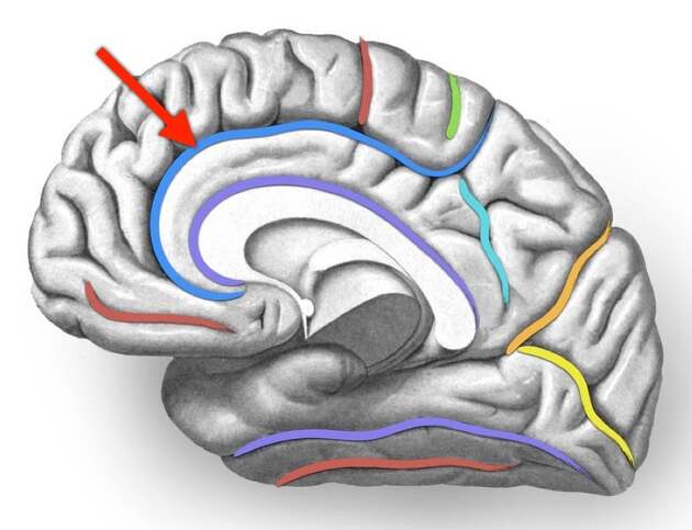
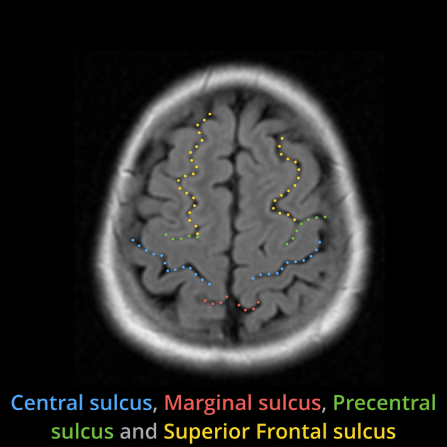
- limiting sulcus: some sulci develop between areas differing in structure and function (e.g. central sulcus)
- axial sulcus: some sulci develop along the axis of a rapidly growing/developing area (e.g. calcarine sulcus)
- operculated sulcus: a sulcus may be between two structurally-different areas and a third sulcus may lie in its wall and does not appear on the surface (e.g. lunate sulcus)
On the basis of formation 3:
- primary sulcus: formed before birth (e.g. central sulcus)
- secondary sulcus: sulci formed within others due to the continued growth of adjoining areas of cortex (e.g. the splenium of the corpus callosum conveys a large number of fibers from the temporal and occipital cortices to the parietal-occipital sulcus creating a number of axial and limiting sulci within the wall of the parieto-occipital sulcus)
On the basis of continuity (whether the sulci are broken into segments by gyral bridges crossing them) 2:
- commonly continuous or uninterrupted (e.g. lateral sulcus, which results from the relatively slow growth of the insular cortex and the submersion of the adjoining frontal, parietal and temporal areas)
- those that have low interruption rates (e.g. central, collateral and calcarine sulci)
- those that are regularly interrupted (e.g. postcentral, superior and inferior frontal sulci)
History and etymology
- sulcus (Latin: furrow, groove)
- fissure (Latin: cleft, groove)
Related Radiopaedia articles
Anatomy: Brain
-
brain
- grey matter
- white matter
-
cerebrum
-
cerebral hemisphere (telencephalon)
- cerebral lobes and gyri
- frontal lobe
- parietal lobe
-
occipital lobe
- occipital pole
- lingual gyrus
- fusiform gyrus (Brodmann area 37)
- calcarine (visual) cortex
- cuneus
- temporal lobe
- basal forebrain
- limbic system
- insula
-
cerebral sulci and fissures (A-Z)
- calcarine fissure
- callosal sulcus
- central (Rolandic) sulcus
- cingulate sulcus
- collateral sulcus
- inferior frontal sulcus
- inferior occipital sulcus
- inferior temporal sulcus
- interhemispheric fissure
- intraparietal sulcus
- lateral (Sylvian) sulcus
- lateral occipital sulcus
- marginal sulcus
- occipitotemporal sulcus
- olfactory sulcus
- paracentral sulcus
- paraolfactory sulcus
- parieto-occipital fissure
- posterior parolfactory sulcus
- precentral sulcus
- preoccipital notch
- postcentral sulcus
- rhinal sulcus
- rostral sulcus
- subparietal sulcus
- superior frontal sulcus
- superior occipital sulcus
- superior temporal sulcus
- cortical histology
- cerebral lobes and gyri
- white matter tracts
- deep grey matter
-
pituitary gland
- posterior pituitary and stalk (part of diencephalon)
- anterior pituitary
- inferior hypophyseal arterial circle
- diencephalon
-
cerebral hemisphere (telencephalon)
-
brainstem
- midbrain (mesencephalon)
- pons (part of metencephalon)
- medulla oblongata (myelencephalon)
- white matter
-
grey matter
- non-cranial nerve
-
cranial nerve nuclei
- oculomotor nucleus
- Edinger-Westphal nucleus
- trochlear nucleus
- motor nucleus of CN V
- mesencephalic nucleus of CN V
- main sensory nucleus of CN V
- spinal nucleus of CN V
- abducent nucleus
- facial nucleus
- superior salivatory nucleus
- cochlear nuclei
- vestibular nuclei
- inferior salivatory nucleus
- solitary tract nucleus
- ambiguus nucleus
- dorsal vagal motor nucleus
- hypoglossal nucleus
-
cerebellum (part of metencephalon)
- vermis
- cerebellar hemisphere
- cerebellar peduncles
- cranial meninges (meninx primitiva)
- CSF spaces
-
cranial nerves (mnemonic)
- olfactory nerve (CN I)
- optic nerve (CN II)
- oculomotor nerve (CN III)
- trochlear nerve (CN IV)
- trigeminal nerve (CN V) (mnemonic)
- abducens nerve (CN VI)
- facial nerve (CN VII) (segments mnemonic | branches mnemonic)
-
vestibulocochlear nerve (CN VIII)
- vestibular ganglion (Scarpa's ganglion)
- glossopharyngeal nerve (CN IX)
- vagus nerve (CN X)
- spinal accessory nerve (CN XI)
- hypoglossal nerve (CN XII)
- functional neuroanatomy
- CNS development
- cerebral vascular supply
- arteries
- vascular territories
-
circle of Willis
- internal carotid artery (ICA) (segments)
- vertebral artery
-
normal variants
- intracranial arterial fenestration
- internal carotid artery (ICA)
- anterior cerebral artery (ACA)
- middle cerebral artery (MCA)
- posterior cerebral artery (PCA)
- basilar artery
- persistent carotid-vertebrobasilar artery anastomoses (mnemonic)
- vertebral artery
- ophthalmic artery
-
cerebral venous system
-
dural venous sinuses
- basilar venous plexus
- cavernous sinus (mnemonic)
- clival diploic veins
- inferior petro-occipital vein
- inferior petrosal sinus
- inferior sagittal sinus
- intercavernous sinus
- internal carotid artery venous plexus of Rektorzik
- jugular bulb
- marginal sinus
- occipital sinus
- sigmoid sinus
- sphenoparietal sinus
- straight sinus
- superior petrosal sinus
- superior sagittal sinus
- torcula herophili
- transverse sinus
-
cerebral veins
-
superficial veins of the brain
- superior cerebral veins (superficial cerebral veins)
- inferior cerebral veins
- superficial middle cerebral vein
- superior anastomotic vein (of Trolard)
- inferior anastomotic vein (of Labbe)
-
superficial veins of the brain
- deep veins of the brain
- normal variants
-
dural venous sinuses
- arteries
- glymphatic pathway