
Chest x-ray: ET tube position (summary)
Citation, DOI, disclosures and article data
Citation:
Jones J, Hacking C, Bell D, et al. Chest x-ray: ET tube position (summary). Reference article, Radiopaedia.org (Accessed on 02 Mar 2025) https://doi.org/10.53347/rID-31404
Permalink:
rID:
31404
Article created:
Disclosures:
At the time the article was created Jeremy Jones had no recorded disclosures.
View Jeremy Jones's current disclosures
Last revised:
Disclosures:
At the time the article was last revised Craig Hacking had no recorded disclosures.
View Craig Hacking's current disclosures
Revisions:
11 times, by
5 contributors -
see full revision history and disclosures
Systems:
Sections:
Tags:
This is a basic article for medical students and other non-radiologists
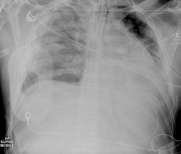
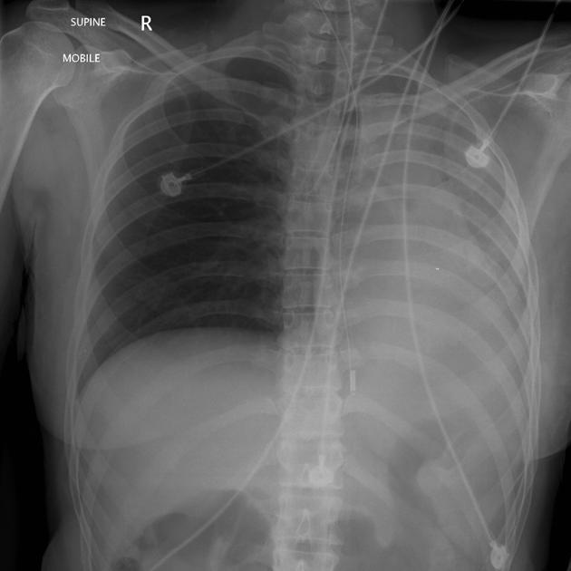
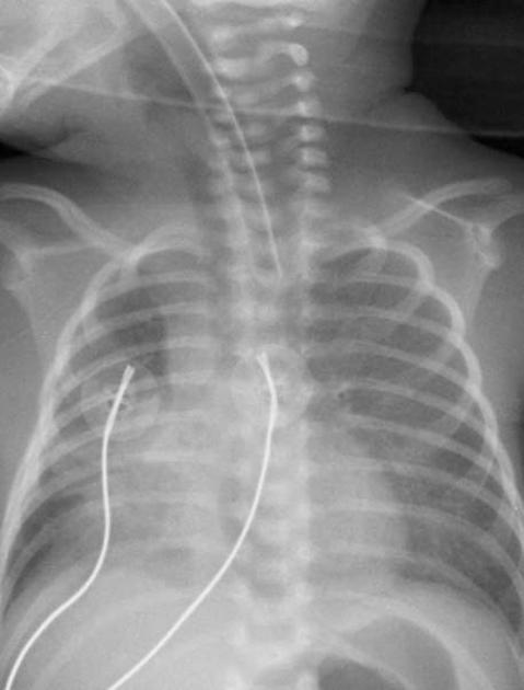
Chest x-ray ET (endotracheal) tube position should be assessed following initial placement and on subsequent radiographs.
Reference article
This is a summary article; we have a more in-depth reference article, see ETT.
Summary
-
normal
- tip 5 cm above carina
- width 2/3 tracheal diameter
- cuff should not expand the trachea
-
malposition
- tip may extend past the carina
- malposition most commonly seen is the tip in the right main bronchus
-
change in position
- ET tube tip position is dependent on head position
- tip position changes depending on neck flexion/extension
- tip position may change by up to 2 cm
- neck flexion moves the tip downwards
- neck extension moves the tip upwards
Incoming Links
Related articles: Education: Medical student curriculum
- radiology for students
-
neuroradiology
- imaging
- key findings
- conditions
- presentations
- cardiac radiology
-
chest radiology
- imaging
- key findings
- conditions
- presentations
-
abdominal radiology
- imaging
- key findings
- conditions
- upper GI
- lower GI
- hepatopancreatobiliary
- genitourinary
- vascular
- breast
- presentations
-
musculoskeletal radiology
- imaging
- key findings
- interpretation
- conditions
- upper limb
- lower limb
- pelvic fractures
- proximal femoral fractures
- distal fibula fracture
- 5th metatarsal fracture
- pediatrics
- spine
- major trauma
- joint pain/arthritis
- presentations
- upper limb
- lower limb
- hip trauma
- lower limb injury
- foot and ankle injury
- joint pain/arthritis
-
obstetrics and gynecology imaging
- imaging
- pelvic US - transabdominal
- pelvic US - transvaginal
- hysterosalpingogram
- CT abdomen
- MRI pelvis
- key findings
- endometrial thickening
- ovarian cysts
- conditions
- non-obstetric
- pelvic inflammatory disease
- tubo-ovarian abscess
- ovarian torsion
- ovarian neoplasms
- endometriosis
- endometrial hyperplasia
- endometrial carcinoma
- cervical cancer
- obstetric
- normal pregnancy
- abnormal first trimester
- ectopic pregnancy
- heterotopic pregnancy
- twins
- non-obstetric
- presentations
- PV bleeding
- pelvic pain
- PV discharge
- early pregnancy
- imaging
-
pediatric radiology
- imaging
- key findings
- conditions
- presentations


 Unable to process the form. Check for errors and try again.