
Ciliary ganglion
Updates to Article Attributes
Body
was changed:
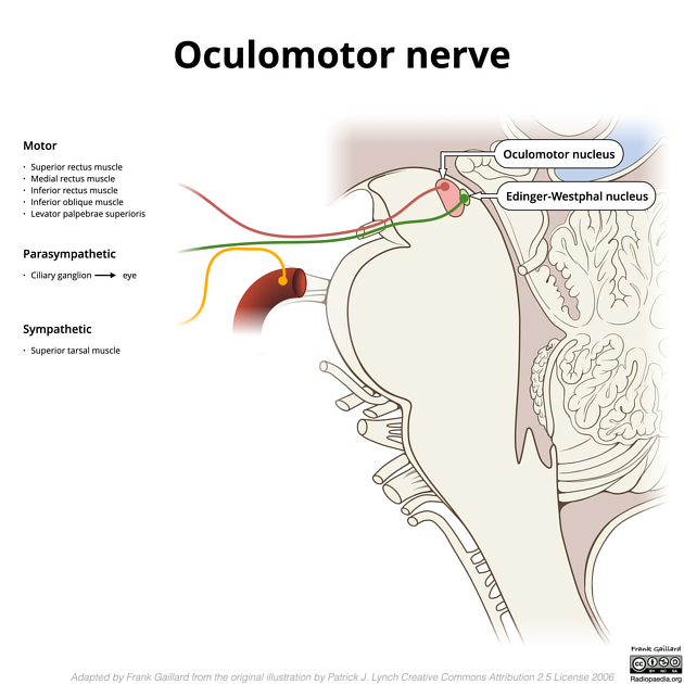
The ciliary ganglion is one of four parasympathetic ganglia of the head and neck. It receives parasympathetic fibres from the oculomotor nerve.
Gross anatomy
- smallest of the ganglia (2mm in size)
- located posterolaterally in the intraconal space of the orbit (towards the orbital apex) between the optic nerve and the lateral rectus muscle
- just lateral to the ophthalmic artery as it crosses the optic nerve from lateral to medial
Roots
- parasympathetic root (fibres synapse in the ganglion)
- from the Edinger-Westphal nucleus of the oculomotor nerve
- sympathetic root (fibres pass through the ganglion without synapsing)
- from the ICA (from the superior cervical ganglion) via the nasociliary nerve
- sensory root (fibres pass through the ganglion without synapsing)
Branches
- short ciliary nerves from the ophthalmic division of the trigeminal nerve which supply the ciliary body (for accommodation) and the sphincter papillae (for pupillary constriction)
Related pathology
-
Pathologypathology of the ciliary ganglion can produce a tonic pupil, where the pupil does not react to light and slowly accommodates - Adie syndrome: when a non-reactive, slowly accomodating pupil is associated with absent deep tendon reflexes and diaphoresis


 Unable to process the form. Check for errors and try again.