
Lateral rectus muscle
Citation, DOI, disclosures and article data
At the time the article was created Jeremy Jones had no recorded disclosures.
View Jeremy Jones's current disclosuresAt the time the article was last revised Craig Hacking had no recorded disclosures.
View Craig Hacking's current disclosures- Lateral rectus
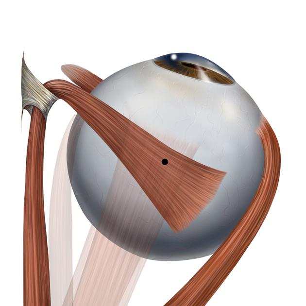
The lateral rectus muscle is one of the six extraocular muscles that control eye movements. It is responsible for abduction and is the only muscle that is innervated by the abducens nerve (CN VI).
On this page:
Summary
- innervation: abducens nerve (CN VI)
- origin: annulus of Zinn (tendinous ring)
- insertion: globe (anterior, lateral surface)
- primary function: one of three ocular abductors
Gross anatomy
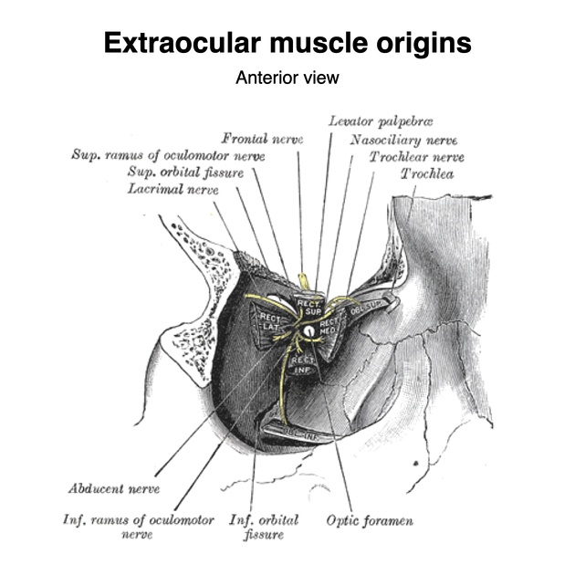
Origin
Lateral rectus, along with the other rectus muscles, arises from the annulus of Zinn, the common tendinous ring at the apex of the orbit that surrounds the optic canal 1.
Insertion
Lateral rectus runs anteriorly on the lateral surface of the eye and inserts into the lateral surface of the sclera just posterior to the junction of cornea and sclera 2.
Arterial supply
Branches of the ophthalmic artery, itself a branch of the internal carotid artery.
Innervation
Lateral rectus is unique among the extraocular muscles in being supplied by the abducens nerve.
Action
The action of the lateral rectus is to abduct the eye (see figure 1) 1. Unlike most of the other extraocular muscles, it has no significant contribution to movement in the other ocular axes.
Etymology
Rectus comes from the Latin rectos, meaning straight 1.
Related pathology
-
oculomotor nerve palsy
- as the lateral rectus and superior oblique muscles are the only extraocular muscles not supplied by the oculomotor nerve, the unopposed force generated by these muscles results in depression and abduction of the affected eye
-
abducens nerve palsy
- as the lateral rectus is the primary ocular abductor, abducens nerve palsy results in adduction of the affected eye, due to unopposed effect primarily of the medial rectus muscle
- because of its long subarachnoid course and location in the Dorello canal, abducens nerve palsy can be an early sign of increased intracranial pressure
See also
References
- 1. Moore KL, Agur AMR, Dalley AF. Clinically Oriented Anatomy. (2013) ISBN: 9781451119459
- 2. Netter FH. Atlas of Human Anatomy. (2018) ISBN: 9780323393225
- 3. Imaging of the Head and Neck. Thieme. (2012) ISBN:3131505311. Read it at Google Books - Find it at Amazon
- 4. Gray's basic anatomy. Churchill Livingstone. ISBN:1455710784. Read it at Google Books - Find it at Amazon
Incoming Links
- IgG4-related orbital disease
- Stability of the eye
- Abducens nerve palsy
- Facial colliculus syndrome
- Extraocular muscle nerve supply (mnemonic)
- Ciliary ganglion
- Myofascial cone
- Ocular abductors
- Orbital apex
- Greater wing of sphenoid
- Ocular globe
- Duane syndrome
- Senile calcific scleral plaques
- Internuclear ophthalmoplegia
- Orbital nerve supply
- Extraocular muscles
- Lacrimal artery
- Barrett's index
- Abducens nerve
- Eye movements
- Sinonasal nonkeratinizing squamous cell carcinoma
- Orbital metastasis of breast cancer
- Cystic lymphangioma of the orbital cavity
- Pleomorphic adenoma of lacrimal gland
- Schwannoma of the orbital cavity
- Basal cell carcinoma of the lateral canthus
- Basal cell carcinoma of the eyelid
- Nonspecific orbital inflammation
- Chondrosarcoma of the maxilla
- Adenoid cystic carcinoma of lacrimal gland
- Spheno-orbital and calvarium intraosseous meningioma
- Lateral orbital wall blow-in fracture and concomitant orbital floor and maxillary sinus wall fracture
- Innervation of the medial and lateral recti muscles (Gray's illustration)
- Orbital cavernous haemangioma
- Orbital pseudotumour
- Orbital foreign body: stiletto shoe
Related articles: Anatomy: Head and neck
- skeleton of the head and neck
-
cranial vault
- scalp (mnemonic)
- fontanelle
-
sutures
- calvarial
- facial
- frontozygomatic suture
- frontomaxillary suture
- frontolacrimal suture
- frontonasal suture
- temporozygomatic suture
- zygomaticomaxillary suture
- parietotemporal suture (parietomastoid suture)
- occipitotemporal suture (occipitomastoid suture)
- sphenofrontal suture
- sphenozygomatic suture
- spheno-occipital suture (not a true suture)
- lacrimomaxillary suture
- nasomaxillary suture
- internasal suture
- basal/internal
- skull landmarks
- frontal bone
- temporal bone
- parietal bone
- occipital bone
- skull base (foramina)
-
facial bones
- midline single bones
- paired bilateral bones
- cervical spine
- hyoid bone
- laryngeal cartilages
-
cranial vault
- muscles of the head and neck
- muscles of the tongue (mnemonic)
- muscles of mastication
-
facial muscles
- epicranius muscle
- circumorbital and palpebral muscles
- nasal muscles
-
buccolabial muscles
- elevators, retractors and evertors of the upper lip
- levator labii superioris alaeque nasalis muscle
- levator labii superioris muscle
- zygomaticus major muscle
- zygomaticus minor muscle
- levator anguli oris muscle
- malaris muscle
- risorius muscle
- depressors, retractors and evertors of the lower lip
- depressor labii inferioris muscle
- depressor anguli oris muscle
- mentalis muscle
- compound sphincter
-
orbicularis oris muscle
- incisivus labii superioris muscle
- incisivus labii inferioris muscle
-
orbicularis oris muscle
- muscle of mastication
- modiolus
- elevators, retractors and evertors of the upper lip
- muscles of the middle ear
- orbital muscles
- muscles of the soft palate
- pharyngeal muscles
- suprahyoid muscles
- infrahyoid muscles
- intrinsic muscles of the larynx
- muscles of the neck
- platysma muscle
- longus colli muscle
- longus capitis muscle
- scalenus anterior muscle
- scalenus medius muscle
- scalenus posterior muscle
- scalenus pleuralis muscle
- sternocleidomastoid muscle
-
suboccipital muscles
- rectus capitis posterior major muscle
- rectus capitis posterior minor muscle
- obliquus capitis superior muscle
- obliquus capitis inferior muscle
- accessory muscles of the neck
- deep cervical fascia
-
deep spaces of the neck
- anterior cervical space
- buccal space
- carotid space
- danger space
- deep cervical fascia
- infratemporal fossa
- masticator space
- parapharyngeal space
- stylomandibular tunnel
- parotid space
- pharyngeal (superficial) mucosal space
- perivertebral space
- posterior cervical space
- pterygopalatine fossa
- retropharyngeal space
- suprasternal space (of Burns)
- visceral space
- surgical triangles of the neck
- orbit
- ear
- paranasal sinuses
- upper respiratory tract
- viscera of the neck
- blood supply of the head and neck
-
arterial supply
-
common carotid artery
- carotid body
- carotid bifurcation
- subclavian artery
- variants
-
common carotid artery
- venous drainage
-
arterial supply
- innervation of the head and neck
-
cranial nerves
- olfactory nerve (CN I)
- optic nerve (CN II)
- oculomotor nerve (CN III)
- trochlear nerve (CN IV)
-
trigeminal nerve (CN V) (mnemonic)
- trigeminal ganglion
- ophthalmic division
- maxillary division
- mandibular division
- abducens nerve (CN VI)
- facial nerve (CN VII)
-
vestibulocochlear nerve (CN VIII)
- vestibular ganglion (Scarpa's ganglion)
- glossopharyngeal nerve (CN IX)
- vagus nerve (CN X)
- (spinal) accessory nerve (CN XI)
- hypoglossal nerve (CN XII)
- parasympathetic ganglia of the head and neck
- cervical sympathetic ganglia
- greater occipital nerve
- third occipital nerve
-
cervical plexus
- muscular branches
- longus capitis
- longus colli
- scalenes
- geniohyoid
- thyrohyoid
-
ansa cervicalis
- omohyoid (superior and inferior bellies separately)
- sternothyroid
- sternohyoid
- phrenic nerve
- contribution to the accessory nerve (CN XI)
- cutaneous branches
- muscular branches
- brachial plexus
- pharyngeal plexus
-
cranial nerves
- lymphatic drainage of the head and neck
- embryological development of the head and neck


 Unable to process the form. Check for errors and try again.