
Ciliary ganglion
- Philips Australia, Paid speaker at Philips Spectral CT events (ongoing)
Updates to Article Attributes
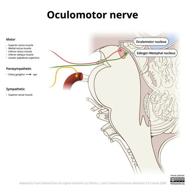
The ciliary ganglion is one of four parasympathetic ganglia of the head and neck. It receives preganglionic parasympathetic fibres from the Edinger-Westphal nucleus via the oculomotor nerve. It supplies the eye via short ciliary nerves not only with parasympathetic fibres, but also with sensory and sympathetic fibres that pass through the ganglion.
Gross anatomy
smallest of the ganglia (2 mm in size)
located posterolaterally in the intraconal space of the orbit (towards the orbital apex) between the optic nerve and the lateral rectus muscle
just lateral to the ophthalmic artery as it crosses the optic nerve from lateral to medial
Roots
Although the ciliary ganglion has parasympathetic, sensory and sympathetic roots, only the parasympathetic ones synapse within the ganglion.
-
parasympathetic root
from the Edinger-Westphal nucleus of the oculomotor nerve (III)
fibres synapse in the ganglion
-
sympathetic root
from the ICA (from the superior cervical ganglion) via the nasociliary nerve, a branch of the trigeminal nerve
fibres pass through the ganglion without synapsing
-
sensory root
via the small communicating branch of the ciliary ganglion (from the nasociliary nerve, a branch of the trigeminal nerve)
fibres pass through the ganglion without synapsing
Branches
-
~12 or more branches, termed
each contains elements from all 3 roots (above), and pierce the back of the sclera around the attachment of the optic nerve to supply the globe
the vast majority of fibres from ganglionic cells supply the ciliary body (accommodation); only ~3% supply sphincter pupillae
Note: while both long ciliary nerves (branches of the nasociliary nerve) and short ciliary nerves contain sensory/sympathetic supply to the cornea, iris, and ciliary body, only the short ciliary nerves are involved in pupillary constriction and accommodation.
Related pathology
pathology of the ciliary ganglion can produce a tonic pupil, where the pupil does not react to light and slowly accommodates
Adie syndrome: when a non-reactive, slowly accomodating pupil is associated with absent deep tendon reflexes and diaphoresis


 Unable to process the form. Check for errors and try again.