
Duodenojejunal flexure
Citation, DOI, disclosures and article data
At the time the article was created Yaïr Glick had no recorded disclosures.
View Yaïr Glick's current disclosuresAt the time the article was last revised Craig Hacking had the following disclosures:
- Philips Australia, Paid speaker at Philips Spectral CT events (ongoing)
These were assessed during peer review and were determined to not be relevant to the changes that were made.
View Craig Hacking's current disclosures- Duodenojejunal junction
- Duodenal-jejunal flexure
- Duodenal-jejunal junction
- DJ flexure
- DJ junction
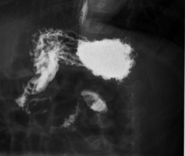
The duodenojejunal (DJ) flexure or junction is the anatomical border between the duodenum and the jejunum.
On this page:
Gross anatomy
The duodenojejunal flexure is located anterolateral to the aorta at the level of the upper border of the second lumbar vertebra. It makes a sharp turn anteroinferiorly to become the jejunum.
The duodenojejunal flexure is suspended from the ligament of Treitz, which serves as its surgical landmark and gives it its unique shape. It also marks the small bowel's transition from the retroperitoneum, which invests the duodenum, into the peritoneal cavity.
Relations
Anterior
- transverse colon and mesocolon
- peritoneum of the root of the small bowel mesentery
Posterior
- aorta
- inferior mesenteric vein (or at its lateral margin)
- left sympathetic trunk
- left renal vessels
- left gonadal vessels
Superior
- inferior border of body of pancreas
Blood supply
Arterial
- branches from the first jejunal branch of the superior mesenteric artery
- frequently, a terminal branch of the anterior superior pancreaticoduodenal artery
Venous
- venous anatomy not well characterized
- veins accompany arteries of corresponding names and may drain directly into the superior mesenteric vein
Innervation
sympathetic: periarterial celiac plexus and superior mesenteric plexus
parasympathetic: vagal fibers from the celiac plexus synapse on neurons in the duodenal wall
Variant anatomy
For uncommon variant anatomy of the duodenojejunal flexure, see
References
- 1. Susan Standring. Gray's Anatomy: The Anatomical Basis of Clinical Practice, 41st edition. Elsevier, 2016. ISBN: 9780702052309
Incoming Links
Related articles: Anatomy: Abdominopelvic
- skeleton of the abdomen and pelvis
- muscles of the abdomen and pelvis
- spaces of the abdomen and pelvis
- anterior abdominal wall
- posterior abdominal wall
- abdominal cavity
- pelvic cavity
- perineum
- abdominal and pelvic viscera
- gastrointestinal tract
- spleen
- hepatobiliary system
-
endocrine system
-
adrenal gland
- adrenal vessels
- chromaffin cells
- variants
- pancreas
- organs of Zuckerkandl
-
adrenal gland
-
urinary system
-
kidney
- renal pelvis
- renal sinus
- avascular plane of Brodel
-
variants
- number
- fusion
- location
- shape
- ureter
- urinary bladder
- urethra
- embryology
-
kidney
- male reproductive system
-
female reproductive system
- vulva
- vagina
- uterus
- adnexa
- Fallopian tubes
- ovaries
- broad ligament (mnemonic)
- variant anatomy
- embryology
- blood supply of the abdomen and pelvis
- arteries
-
abdominal aorta
- inferior phrenic artery
- celiac artery
- superior mesenteric artery
- middle suprarenal artery
- renal artery (variant anatomy)
- gonadal artery (ovarian artery | testicular artery)
- inferior mesenteric artery
- lumbar arteries
- median sacral artery
-
common iliac artery
- external iliac artery
-
internal iliac artery (mnemonic)
- anterior division
- umbilical artery
- superior vesical artery
- obturator artery
- vaginal artery
- inferior vesical artery
- uterine artery
- middle rectal artery
-
internal pudendal artery
- inferior rectal artery
-
perineal artery
- posterior scrotal artery
- transverse perineal artery
- artery to the bulb
- deep artery of the penis/clitoris
- dorsal artery of the penis/clitoris
- inferior gluteal artery
- posterior division (mnemonic)
- variant anatomy
- anterior division
-
abdominal aorta
- portal venous system
- veins
- anastomoses
- arterioarterial anastomoses
- portal-systemic venous collateral pathways
- watershed areas
- arteries
- lymphatics
- innervation of the abdomen and pelvis
- thoracic splanchnic nerves
- lumbar plexus
-
sacral plexus
- lumbosacral trunk
- sciatic nerve
- superior gluteal nerve
- inferior gluteal nerve
- nerve to piriformis
- perforating cutaneous nerve
- posterior femoral cutaneous nerve
- parasympathetic pelvic splanchnic nerves
- pudendal nerve
- nerve to quadratus femoris and inferior gemellus muscles
- nerve to internal obturator and superior gemellus muscles
- autonomic ganglia and plexuses


 Unable to process the form. Check for errors and try again.