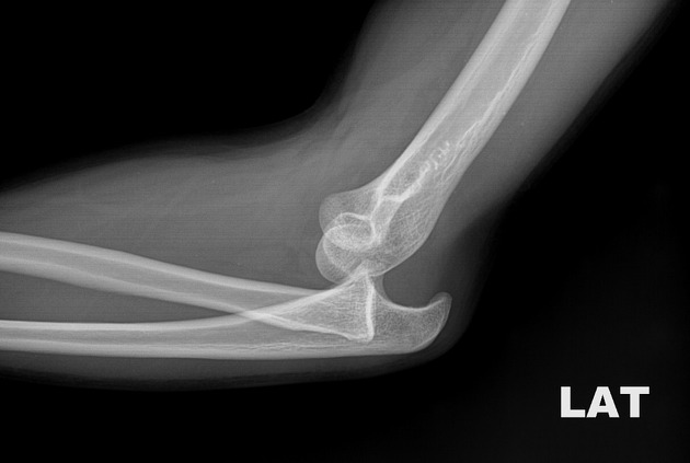
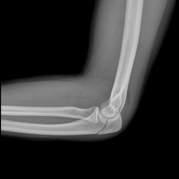
This is a basic article for medical students and other non-radiologists
Elbow radiographs are common plain films that are obtained frequently in the emergency department.
Summary approach
-
alignment
-
drawn down the anterior surface of the humerus
should intersect the middle 1/3 of the capitellum
if it does not, think distal humeral fracture
-
drawn along the radial neck
should always intersect the capitellum
-
if it does not, think radial head dislocation or subluxation
check for an accompanying fracture, e.g. Monteggia fracture-dislocation
-
-
effusion
visible posterior fat pad always indicates an elbow effusion
-
if there is an effusion, think acute intra-articular fracture
-
elbow fractures may be occult on x-rays
adult: radial head fracture
child: supracondylar fracture
-
-
small anterior fat pad may be seen in normal patients
only significant if massively raised
-
bones
-
look specifically for common fractures
radial head and neck fractures
olecranon fracture
-
-
cortex
-
trace the cortex of each bone
distal humerus
radial head, neck and shaft
olecranon, coronoid process and ulnar shaft
-


 Unable to process the form. Check for errors and try again.