
Ethmoid bone
Citation, DOI, disclosures and article data
At the time the article was created Jeremy Jones had no recorded disclosures.
View Jeremy Jones's current disclosuresAt the time the article was last revised Frank Gaillard had no financial relationships to ineligible companies to disclose.
View Frank Gaillard's current disclosures- Ethmoidal bone
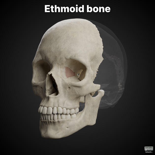
The ethmoid bone is a single midline facial bone that separates the nasal cavity from the brain and is located at the roof of the nose and between the orbits. It is a cubical shape and is relatively lightweight because of its spongy construction and air-filled sinuses. It contributes to the anterior cranial fossa.
Gross anatomy
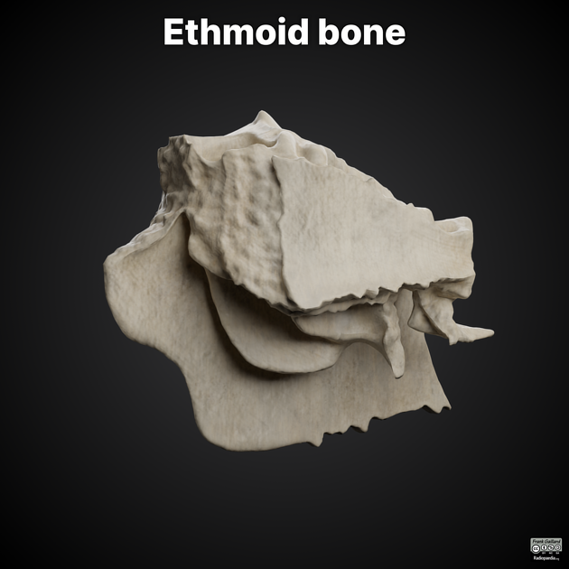
The ethmoid bone consists of four parts:
horizontal (cribriform) plate
vertical (perpendicular) plate
2 lateral masses (labyrinths)
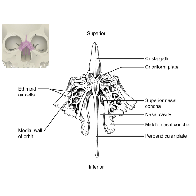
The horizontal plate features include:
foramina in cribriform plate: for olfactory nerve filaments (20), olfactory bulb lies in the olfactory fossa
crista galli (L: rooster's crest): thick, smooth triangular process, attachment for falx cerebri
nasal slits: anteriorly, passage of anterior ethmoidal nerve
The vertical plate forms the part of the bony nasal septum:
anterior border articulates with the frontal bone and crests of nasal bones
posterior border features the sphenoidal crest which articulates with the vomer
Labyrinths (2) house anterior and posterior ethmoidal air cells.
lateral surface features include: the orbit, lamina papyracea, uncinate process below: medial wall of the maxillary sinus which articulates with the inferior concha
medial surface features include: the nasal cavity, superior nasal concha, and middle nasal concha helps form the middle meatus, and the infundibulum opens there
upper surface: grooves articulate with frontal bone to form anterior and posterior ethmoidal canals
Articulations
Articulates with 15 bones in total (4 cranial bones and 11 facial bones):
References
- 1. Henry Gray, Patricia Collins. Gray's Anatomy. (2005) ISBN: 0443071683 - Google Books
Incoming Links
- Olfactory nerve
- Maxilla
- Ethmoidal air cells
- Craniofacial fibrous dysplasia
- Keros classification of olfactory fossa
- Olfactory fossa
- Base of the skull
- Uncinate process
- Lamina papyracea
- Olfactory system
- Nasal septum
- Skull vault bones (mnemonic)
- Vomer
- Axial skeleton
- Middle nasal concha
- Crista galli
- Cranial vault
- Nasal septal cartilage
- Frontal bone
- Palatine bone
- Internal angular orbital dermoid cyst - fluid density
- Frontoethmoidal sinus osteoma
- Multifocal orbital and calvarial fractures
- Ocular rupture and orbital blow-out fracture
- Ethmoid sinus osteoma
- Orbital roof fracture and intracerebral hemorrhage
- Mucocele of the ethmoidal sinus
- Ethmoid bone (Gray's illustrations)
- Nasal cavity (Gray's illustration)
Related articles: Anatomy: Head and neck
- skeleton of the head and neck
-
cranial vault
- scalp (mnemonic)
- fontanelle
-
sutures
- calvarial
- facial
- frontozygomatic suture
- frontomaxillary suture
- frontolacrimal suture
- frontonasal suture
- temporozygomatic suture
- zygomaticomaxillary suture
- parietotemporal suture (parietomastoid suture)
- occipitotemporal suture (occipitomastoid suture)
- sphenofrontal suture
- sphenozygomatic suture
- spheno-occipital suture (not a true suture)
- lacrimomaxillary suture
- nasomaxillary suture
- internasal suture
- basal/internal
- skull landmarks
- frontal bone
- temporal bone
- parietal bone
- occipital bone
- skull base (foramina)
-
facial bones
- midline single bones
- paired bilateral bones
- cervical spine
- hyoid bone
- laryngeal cartilages
-
cranial vault
- muscles of the head and neck
- muscles of the tongue (mnemonic)
- muscles of mastication
-
facial muscles
- epicranius muscle
- circumorbital and palpebral muscles
- nasal muscles
-
buccolabial muscles
- elevators, retractors and evertors of the upper lip
- levator labii superioris alaeque nasalis muscle
- levator labii superioris muscle
- zygomaticus major muscle
- zygomaticus minor muscle
- levator anguli oris muscle
- malaris muscle
- risorius muscle
- depressors, retractors and evertors of the lower lip
- depressor labii inferioris muscle
- depressor anguli oris muscle
- mentalis muscle
- compound sphincter
-
orbicularis oris muscle
- incisivus labii superioris muscle
- incisivus labii inferioris muscle
-
orbicularis oris muscle
- muscle of mastication
- modiolus
- elevators, retractors and evertors of the upper lip
- muscles of the middle ear
- orbital muscles
- muscles of the soft palate
- pharyngeal muscles
- suprahyoid muscles
- infrahyoid muscles
- intrinsic muscles of the larynx
- muscles of the neck
- platysma muscle
- longus colli muscle
- longus capitis muscle
- scalenus anterior muscle
- scalenus medius muscle
- scalenus posterior muscle
- scalenus pleuralis muscle
- sternocleidomastoid muscle
-
suboccipital muscles
- rectus capitis posterior major muscle
- rectus capitis posterior minor muscle
- obliquus capitis superior muscle
- obliquus capitis inferior muscle
- accessory muscles of the neck
- deep cervical fascia
-
deep spaces of the neck
- anterior cervical space
- buccal space
- carotid space
- danger space
- deep cervical fascia
- infratemporal fossa
- masticator space
- parapharyngeal space
- stylomandibular tunnel
- parotid space
- pharyngeal (superficial) mucosal space
- perivertebral space
- posterior cervical space
- pterygopalatine fossa
- retropharyngeal space
- suprasternal space (of Burns)
- visceral space
- surgical triangles of the neck
- orbit
- ear
- paranasal sinuses
- upper respiratory tract
- viscera of the neck
- blood supply of the head and neck
-
arterial supply
-
common carotid artery
- carotid body
- carotid bifurcation
- subclavian artery
- variants
-
common carotid artery
- venous drainage
-
arterial supply
- innervation of the head and neck
-
cranial nerves
- olfactory nerve (CN I)
- optic nerve (CN II)
- oculomotor nerve (CN III)
- trochlear nerve (CN IV)
-
trigeminal nerve (CN V) (mnemonic)
- trigeminal ganglion
- ophthalmic division
- maxillary division
- mandibular division
- abducens nerve (CN VI)
- facial nerve (CN VII)
-
vestibulocochlear nerve (CN VIII)
- vestibular ganglion (Scarpa's ganglion)
- glossopharyngeal nerve (CN IX)
- vagus nerve (CN X)
- (spinal) accessory nerve (CN XI)
- hypoglossal nerve (CN XII)
- parasympathetic ganglia of the head and neck
- cervical sympathetic ganglia
- greater occipital nerve
- third occipital nerve
-
cervical plexus
- muscular branches
- longus capitis
- longus colli
- scalenes
- geniohyoid
- thyrohyoid
-
ansa cervicalis
- omohyoid (superior and inferior bellies separately)
- sternothyroid
- sternohyoid
- phrenic nerve
- contribution to the accessory nerve (CN XI)
- cutaneous branches
- muscular branches
- brachial plexus
- pharyngeal plexus
-
cranial nerves
- lymphatic drainage of the head and neck
- embryological development of the head and neck


 Unable to process the form. Check for errors and try again.