
Lipomatosis of nerve, also known as fibrolipomatous hamartoma of the nerve, lipofibromatous hamartoma, or neural fibrolipoma, is a benign neoplasm of nerves, resulting from anomalous growth of fibroadipose tissue contained by epineurium resulting in expansion of the nerve with separation of nerve fascicles.
On this page:
Terminology
When neural enlargement results in visible enlargement and deformity of the body, most commonly the hand, the term macrodystrophia lipomatosa is often used, although the conditions are synonymous 9.
Diagnosis
Diagnostic criteria according to the WHO classification of soft tissue and bone tumors (5th edition) 9:
essential: coaxial cable–like appearance on cross-sectional imaging; epineurial expansion by mature adipose tissue with widely spaced nerve fascicles; perineurial thickening and pseudo–onion bulb–like hypertrophic change
Epidemiology
Lipomatosis of nerve is most commonly encountered in children before age 10 years of age 9.
Clinical presentation
This condition either presents as a slowly growing asymptomatic mass or with pain and paresthesia (most common), particularly if associated with nerve compression or entrapment. For example, carpal tunnel syndrome is common when the median nerve is involved in the carpal region 9.
When digital branches of nerves are involved, this can result in macrodactyly of the involved digit (see: macrodystrophia lipomatosa).
Pathology
Lipomatosis of nerve is characterized by hamartomatous overgrowth of mature fat and fibroblasts within the epineurium resulting in marked expansion of the nerve 3,9. Other than case reports with associated proteus syndrome and Klippel-Trénaunay syndrome, there are no known syndromic associations and the condition appears sporadic 9. The WHO classification of tumors of soft tissue categorizes this entity under adipocytic tumors 3.
Location
Lipomatosis of nerve is almost exclusively seen in the limbs, most commonly in the upper limb 9. Only rarely is bilateral or found elsewhere.
-
upper limb: 74%.
median nerve: >60%
ulnar nerve: 7%
-
lower limb: 17%
plantar nerve: 11%
Radiographic features
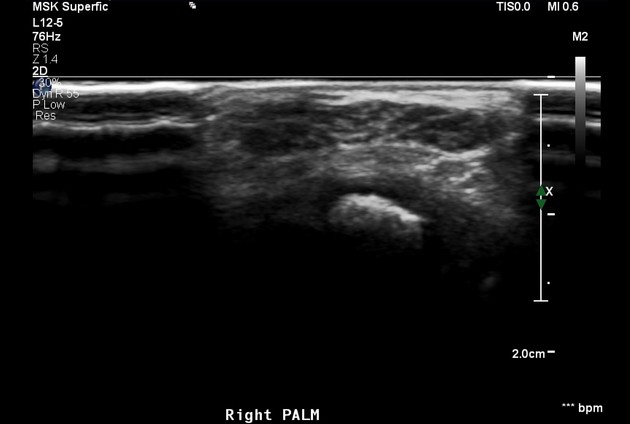
Ultrasound
A characteristic hypoechoic coaxial cabling encased by an echogenic substratum may be seen 9.
MRI
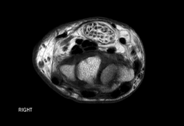
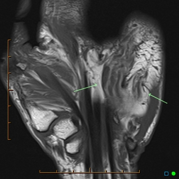
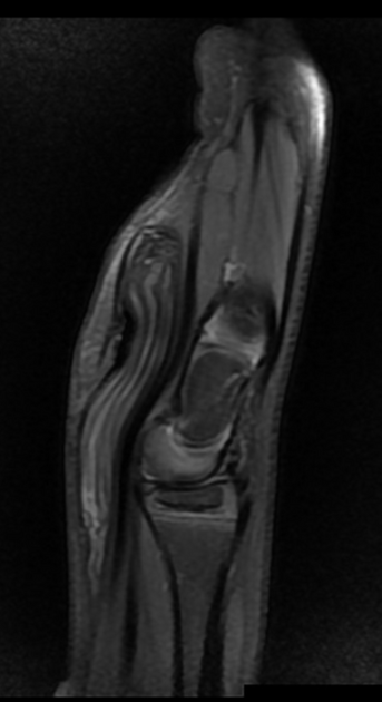
MRI features are often pathognomonic and typically shows a coaxial cable-like appearance on axial images and a spaghetti-like appearance on coronal images 2,6. A pseudo-union-bulb appearance may be present 9.
T1: the neural bundles are hyperintense to muscle and the surrounding substratum is isointense to muscle 3
T2: fat components are high signal and fibrous components are low signal
History and etymology
It is thought to have been first reported by M L Mason in 1953 5.
Treatment and prognosis
Lipomatosis of nerve is benign and therefore treatment is for symptoms but needs to be balanced with preservation of nerve function as it is difficult to resect the lesion without also resecting/impinging the affected nerve.


 Unable to process the form. Check for errors and try again.