
Internal supravesical hernias (alternative plural: herniae) are a type of internal hernia in which viscera protrude into the supravesical fossa, occupying the paravesical space.
On this page:
Epidemiology
It is a very rare condition and accounts for less than 4% of all internal herniae 4.
Clinical presentation
Patients may complain of:
symptoms of small bowel obstruction including abdominal pain, nausea, vomiting, and meteorism
groin pain
urinary symptoms
Pathology
Different types of internal supravesical hernias are described, depending on the location of the hernial sac 3:
prevesical: anterior supravesical (also known as the retropubic space)
paravesical: right/left lateral supravesical
retrovesical: posterior supravesical
Radiographic features
Plain radiograph
Abdominal radiographs demonstrate non-specific signs of small bowel obstruction with:
small bowel loops dilatation
gas-fluid levels
pneumoperitoneum if complicated by perforation
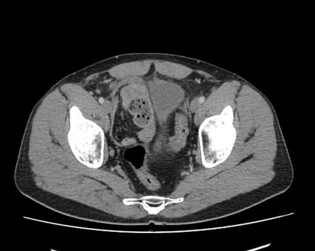
CT
CT is the modality of choice for diagnosis and typical features are:
dilatation of small bowel loops with a transition area in a “closed-loop” at the level of the supravesical fossa
displacement or compression of the lateral wall of the bladder
pneumoperitoneum if complicated with perforation
Treatment and prognosis
Internal supravesical hernias require emergency surgical treatment with laparotomy/laparoscopic procedures.
If the incarcerated loops are viable:
reduction of the incarcerated sac
closure of the defect in the prevesical fascia
If the bowel loops are gangrenous or present doubtful viability:
resection of the bowel loop
end-to-end anastomosis
closure of the defect
Complications
Delay in diagnosis and treatment may lead to intestinal ischemia, perforation and/or peritonitis.
History and etymology
The first case of internal supravesical hernia was reported in 1814 1.


 Unable to process the form. Check for errors and try again.