
This is a basic article for medical students and other non-radiologists
Polyarthritis (arthritis affecting several joints) is common, especially in older patients. Symptoms may range from mild pain and restriction to severe, debilitating disease with mutilated joints.
On this page:
Assessment
Arthritis may be broadly split into degenerative and inflammatory types. The distinction between the two is often possible by taking a detailed history and examining the joints.
Investigations
Further investigation of polyarthritis aims to confirm the diagnosis and assess severity. In both degenerative and inflammatory disease, x-rays are helpful to assess the involvement of the joint surfaces and the condition of the adjacent bones.
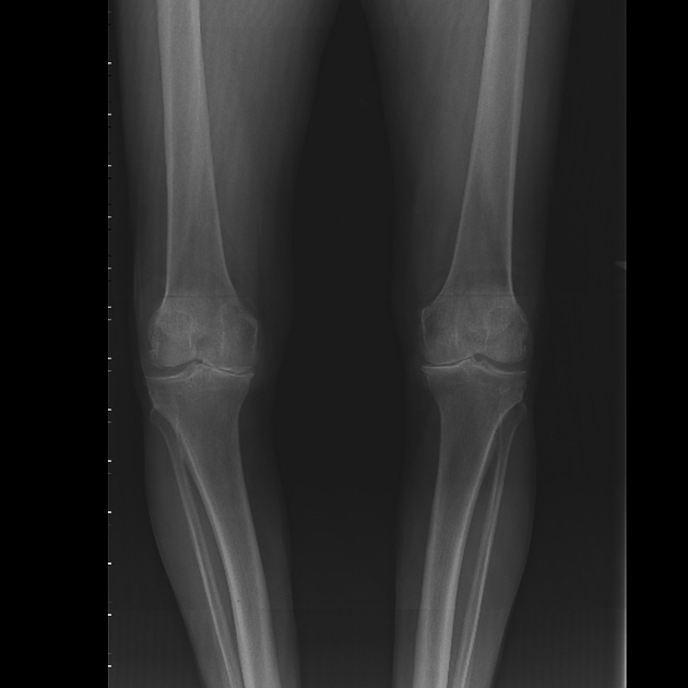
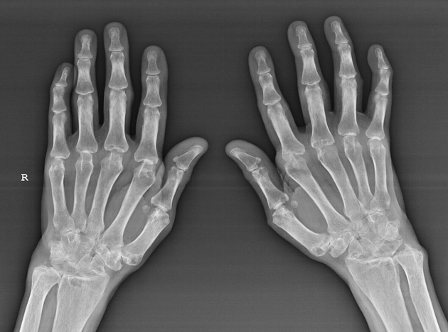
Radiological assessment in chronic joint disease and specifically in polyarthritis often employs x-rays of both sides to allow for comparison.
Degenerative polyarthritis
Degenerative polyarthritis (osteoarthritis) most commonly affects the weight-bearing joints and are commonly seen on pelvic x-rays and AP x-rays of the knees. Common findings include joint space reduction, joint surface sclerosis and irregularity.
Inflammatory polyarthritis
Inflammatory polyarthritis is a broad group of diseases that includes rheumatoid arthritis. Is much less common in the weight-bearing joints and is much more commonly seen in the hands and feet. Inflammatory arthritis also results in joint space reduction but may cause erosions around the joint which is a feature of disease that will trigger escalation of treatment.


 Unable to process the form. Check for errors and try again.