
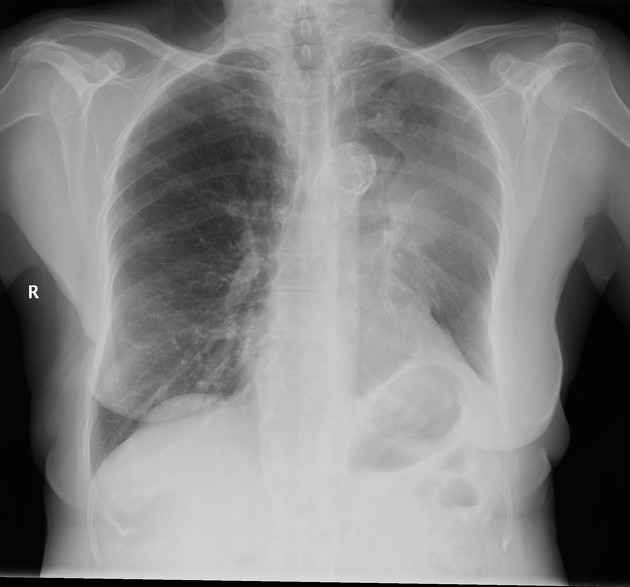
The luftsichel sign is seen in some cases of left upper lobe collapse and refers to the frontal chest radiographic appearance due to hyperinflation of the superior segment of the left lower lobe interposing itself between the mediastinum and the collapsed left upper lobe.
On this page:
Radiographic features
Plain radiograph
In many cases of left upper lobe collapse, the anterior parts of the aortic arch, and thus the aortic knuckle, are abutted by the collapsed lung, and thus, the normal silhouette is lost. In some cases, the apical (superior) segment of the left lower lobe is hyperinflated and becomes interposed between the collapsed lung and the adjacent aortic arch.
In these circumstances, the aortic knuckle silhouette remains visible due to a hyperlucency extending from the apical segment to the superior pulmonary vein 2. The collapsed left upper lobe is thus displaced laterally away from the mediastinum.
CT
CT correlation sometimes reveals that the luftsichel sign is in fact due to fat along the lateral aspect of the aortic arch. This is presumably displaced mediastinal fat.
History and etymology
It is derived from the German words "luft" meaning air, and "sichel" meaning sickle, and literally describes an ‘air crescent’ which may be seen between the aortic arch and the medial border of the collapsed lung.
Differential diagnosis
right lung herniation
medial pneumothorax


 Unable to process the form. Check for errors and try again.